. 2012 Feb;33(4):666-74.
doi: 10.1002/elps.201100434.

A neurosteroid analogue photolabeling reagent labels the colchicine-binding site on tubulin: a mass spectrometric analysis

Affiliations

A neurosteroid analogue photolabeling reagent labels the colchicine-binding site on tubulin: a mass spectrometric analysis

Zi-Wei Chen et al. Electrophoresis. 2012 Feb.

Abstract

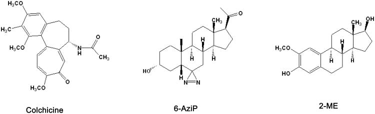
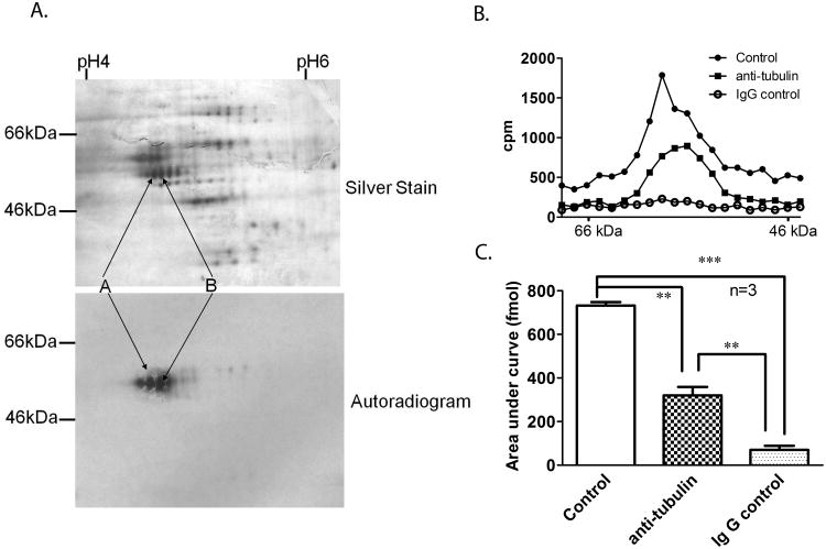
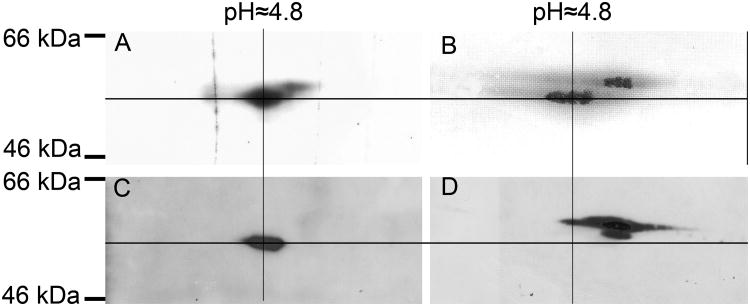
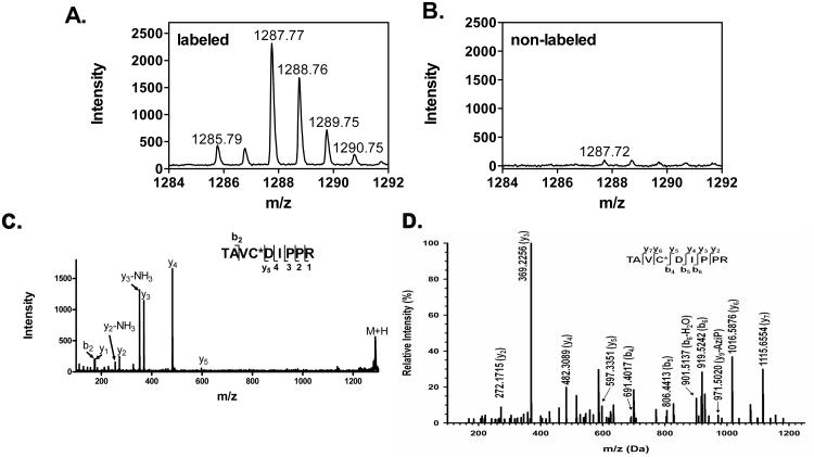
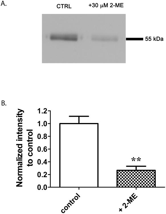
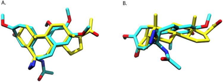
Previous studies have shown that the neurosteroid analogue, 6-Azi-pregnanolone (6-AziP), photolabels voltage-dependent anion channels and proteins of approximately 55 kDa in rat brain membranes. The present study used two-dimensional electrophoresis and nanoelectrospray ionization ion-trap mass spectrometry (nano-ESI-MS) to identify the 55 kDa proteins (isoelectric point 4.8) as isoforms of β-tubulin. This identification was confirmed by immunoblot and immunoprecipitation of photolabeled protein with anti-β-tubulin antibody and by the demonstration that 6-AziP photolabels purified bovine brain tubulin in a concentration-dependent pattern. To identify the photolabeling sites, purified bovine brain tubulin was photolabeled with 6-AziP, digested with trypsin, and analyzed by matrix-assisted laser desorption/ionization MS (MALDI). A 6-AziP adduct of TAVCDIPPR(m/z = 1287.77), a β-tubulin specific peptide, was detected by MALDI. High-resolution liquid chromatography-MS/MS analysis identified that 6-AziP was covalently bound to cysteine 354 (Cys-354), previously identified as a colchicine-binding site. 6-AziP photolabeling was inhibited by 2-methoxyestradiol, an endogenous derivative of estradiol thought to bind to the colchicine site. Structural modeling predicted that neurosteroids could dock in this colchicine site at the interface between α- and β-tubulin with the photolabeling group of 6-AziP positioned proximate to Cys-354.


Figures

Figure 1
Figure 1
Structures of 6-AziP, 2-methoxyestradiol, and colchicine.
Figure 2
Figure 2. Identification of the 55 kDa protein photolabeled by 6-AziP as tubulin
A. Rat brain membranes (250 μg) were photolabeled with 10 μM non-tritiated or tritiated 6-AziP. These two samples were separated by 2-dimensional electrophoresis with both isoelectric focusing and SDS-PAGE being run simultaneously under the same conditions followed by either silver staining (upper panel, for non-tritiated 6-AziP) or autoradiography (lower panel, for [3H]6-AziP). A train of proteins observed in silver staining, corresponding to the radioactive spots (labeled as A and B), were excised from the gel, digested with trypsin and identified as isoforms of α- and β-tubulin using mass spectrometry. B. A representative gel slicing experiment illustrating that most of the photolabeled 55 kDa protein is immunoprecipitated by anti β-tubulin antibody. Rat brain membranes photolabeled with 10 μM [3H]6-AziP were immunoprecipitated with anti β-tubulin antibody or anti-mouse IgG, followed by SDS-PAGE and gel slicing. The control is photolabeled rat brain membrane without immunoprecipitation. Gel slices were analyzed by scintillation spectrometry. C. The areas under the curve of the 55 kDa proteins from 3 replicate gel slicing experiments. They are: 733 ± 15 fmol for control membranes; 321 ± 39 fmol for the anti-β-tubulin immunoprecipitation group, and 71± 19 fmol for the IgG immunoprecipitation group.
Figure 3
Figure 3. 6-AziP preferentially photolabels the β- subunit of tubulin
Purified bovine brain tubulin (10 μg) was photolabeled with either 3 μM [3H]6-AziP or non-tritiated 6-AziP and analyzed by 2-DE. (A). Autoradiogram demonstrating that [3H]6-AziP labels proteins at ≈55 kDa and pI 4.8. (B). Silver staining showing 2 trains of spots representing α- and β-tubulin. (C). Immuno-blot with anti-β-tubulin antibody. (D). Immuno-blot with anti-α-tubulin antibody. Cross marking and alignment of all the gels based on MW and pI indicates that β-tubulin is preferentially photolabeled by 6-AziP.
Figure 4
Figure 4. Mass spectrometry identifies Cys-354 of β-tubulin as the site of 6-AziP incorporation
Comparison of the MALDI mass spectra of photolabeled (A) and non-labeled tubulin (B), identified a peptide with m/z =1287.77 as unique to the photolabeled sample. (C). MALDI tandem mass spectrum identified the peptide as TAVCDIPPR bearing a single 6-AziP adduct. (D). A high-resolution MS2 spectrum (OrbiTrap) demonstrated Cys-354 as the site of 6-AziP photo-incorporation in the peptide TAVC*DIPPR.
Figure 5
Figure 5. 2-ME prevents 6-AziP photolabeling of bovine brain tubulin
10 μg of purified bovine brain tubulin was photolabeled with 0.3 μM [3H]6-AziP in the presence or absence of 2-ME (30 μM). (A). A representative autoradiogram demonstrating that 6-AziP photolabeling is inhibited by 2-ME. (B) Densitometric analysis from 5 replicate experiments indicates that 2-ME prevents 6-AziP photolabeling of bovine brain tubulin by about 75% (student's t-test, **p<0.01). .
Figure 6
Figure 6. Structural similarity of 6-AziP and colchicine
A top (A) and side view (B) of 6-AziP and colchicine. The two superimposed molecules represent the maximal similarity alignment of 6-AziP and colchicine as determined by the program ROCS. In both molecules hydrogens are removed for clarity, the carbons of 6-AziP are depicted in yellow and colchicine in cyan, with oxygens in red and nitrogens in blue. Colchicine and 6-AziP have similar molecular volumes with 6-AziP occupying 339.82 Å3 while colchicine occupies 350.82 Å3.

References

    1. Smith SS. Neurosteroid effects in the central nervous system: the role of the GABAA receptor. CRC Press LLC; 2004.
    1. Gasior M, Carter RB, Witkin JM. Trends Pharmacol Sci. 1999;20:107–112. - PubMed
    1. Rupprecht R. Psychoneuroendocrinology. 2003;28:139–168. - PubMed
    1. Ekins S, Reschly EJ, Hagey LR, Krasowski MD. BMC Evol Biol. 2008;8:103. - PMC - PubMed
    1. Hosie AM, Wilkins ME, da Silva HM, Smart TG. Nature. 2006;444:486–489. - PubMed

MeSH terms

LinkOut - more resources