Neuron deficit in the white matter and subplate in periventricular leukomalacia
- PMID: 22451205
- PMCID: PMC3315053
- DOI: 10.1002/ana.22612
Neuron deficit in the white matter and subplate in periventricular leukomalacia
Abstract
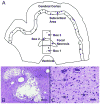
Objective: The cellular basis of cognitive abnormalities in preterm infants with periventricular leukomalacia (PVL) is uncertain. One important possibility is that damage to white matter and subplate neurons that are critical to the formation of the cerebral cortex occurs in conjunction with oligodendrocyte and axonal injury in PVL. We tested the hypothesis that the overall density of neurons in the white matter and subplate region is significantly lower in PVL cases compared to non-PVL controls.
Methods: We used a computer-based method for the determination of the density of microtubule-associated protein 2-immunolabeled neurons in the ventricular/subventricular region, periventricular white matter, central white matter, and subplate region in PVL cases and controls.
Results: There were 5 subtypes of subcortical neurons: granular, unipolar, bipolar, inverted pyramidal, and multipolar. The neuronal density of the granular neurons in each of the 4 regions was 54 to 80% lower (p≤0.01) in the PVL cases (n=15) compared to controls adjusted for age and postmortem interval (n=10). The overall densities of unipolar, bipolar, multipolar, and inverted pyramidal neurons did not differ significantly between the PVL cases and controls. No granular neurons expressed markers of neuronal and glial immaturity (Tuj1, doublecortin, or NG2).
Interpretation: These data suggest that quantitative deficits in susceptible granular neurons occur in the white matter distant from periventricular foci, including the subplate region, in PVL, and may contribute to abnormal cortical formation and cognitive dysfunction in preterm survivors.
Copyright © 2012 American Neurological Association.
Figures


References
-
- Woodward LJ, Moor S, Hood KM, et al. Very preterm children show impairments across multiple neurodevelopmental domains by age 4 years. Arch Dis Child Fetal Neonatal Ed. 2009;94:F339–344. - PubMed
-
- Kinney HC, Volpe JJ. Perinatal panencephalopathy in premature infants: Is it due to hypoxia-ischemia? In: Haddad GG, Yu SP, editors. Brain hypoxia and ischemia with special emphasis on development. New York: Humana Press; 2009. pp. 153–186.
-
- Allendoerfer KL, Shatz CJ. The subplate, a transient neocortical structure: its role in the development of connections between thalamus and cortex. Annu Rev Neurosci. 1994;17:185–218. - PubMed
Publication types
MeSH terms
Grants and funding
LinkOut - more resources
Full Text Sources

