. 2012 May 10;119(19):4408-18.
doi: 10.1182/blood-2011-12-397091. Epub 2012 Mar 26.

Runx1 deletion or dominant inhibition reduces Cebpa transcription via conserved promoter and distal enhancer sites to favor monopoiesis over granulopoiesis

Affiliations

Runx1 deletion or dominant inhibition reduces Cebpa transcription via conserved promoter and distal enhancer sites to favor monopoiesis over granulopoiesis

Hong Guo et al. Blood. .

Abstract

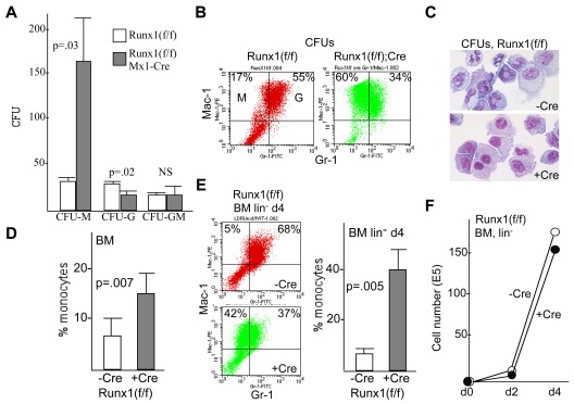
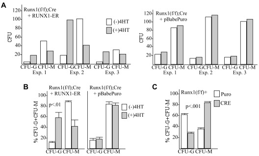
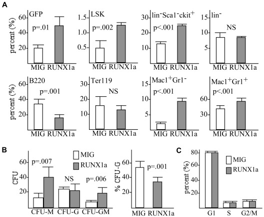
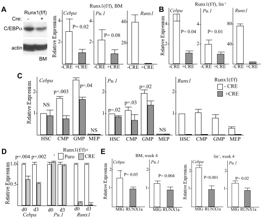
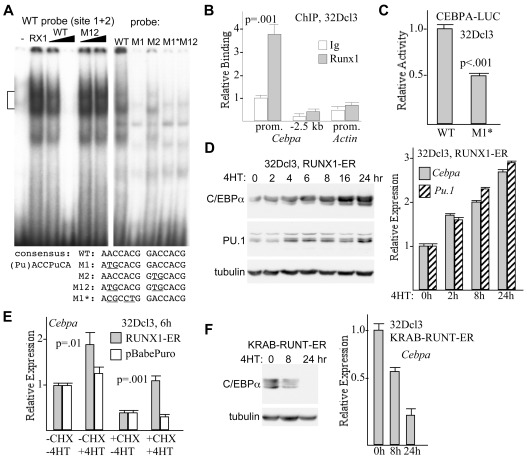
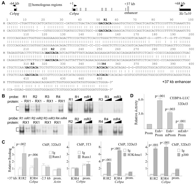
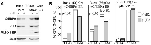
Deletion of Runx1 in adult mice produces a myeloproliferative phenotype. We now find that Runx1 gene deletion increases marrow monocyte while reducing granulocyte progenitors and that exogenous RUNX1 rescues granulopoiesis. Deletion of Runx1 reduces Cebpa mRNA in lineage-negative marrow cells and in granulocyte-monocyte progenitors or common myeloid progenitors. Pu.1 mRNA is also decreased, but to a lesser extent. We also transduced marrow with dominant-inhibitory RUNX1a. As with Runx1 gene deletion, RUNX1a expands lineage-Sca-1+c-kit+ and myeloid cells, increased monocyte CFUs relative to granulocyte CFUs, and reduced Cebpa mRNA. Runx1 binds a conserved site in the Cebpa promoter and binds 4 sites in a conserved 450-bp region located at +37 kb; mutation of the enhancer sites reduces activity 6-fold in 32Dcl3 myeloid cells. Endogenous Runx1 binds the promoter and putative +37 kb enhancer as assessed by ChIP, and RUNX1-ER rapidly induces Cebpa mRNA in these cells, even in cycloheximide, consistent with direct gene regulation. The +37 kb region contains strong H3K4me1 histone modification and p300-binding, as often seen with enhancers. Finally, exogenous C/EBPα increases granulocyte relative to monocyte progenitors in Runx1-deleted marrow cells. Diminished CEBPA transcription and consequent impairment of myeloid differentiation may contribute to leukemic transformation in acute myeloid leukemia cases associated with decreased RUNX1 activity.


Figures

Figure 1
Figure 1
Absence of Runx1 favors monopoiesis over granulopoiesis. (A) Marrow mononuclear cells from Runx1(f/f) or Runx1(f/f);Mx1-Cre mice exposed to pIpC were assessed for myeloid progenitor numbers in IL-3, IL-6, and SCF per 1E4 mononuclear cells plated (mean and SE from 3 determinations). (B) Pooled CFUs were subjected to FACS analysis for Mac-1 and Gr-1. The proportions of Mac-1+Gr-1 monocytes (M) and Mac-1+Gr-1+ granulocytes (G) are indicated. (C) CFUs were cytospun and subjected to Wright-Giemsa staining. (D) Marrow mononuclear cells from Runx1(f/f) or Runx1(f/f);Mx1-Cre mice exposed to pIpC were subjected to similar FACS analysis immediately after isolation. The proportion of Mac-1+Gr-1 monocytes relative to the sum of monocytes and granulocytes is shown (mean and SE from 6 determinations). (E) Control or Runx1-deleted marrow cells were lineage-depleted and placed in liquid culture with IL-3, IL-6, and SCF and subjected to FACS analysis 4 days later. Representative data (left panels) and a summary of 3 determinations (right graph) are shown. (F) Viable cell numbers were enumerated immediately after lineage depletion of control or Runx1-deleted cells and 2 or 4 days later. A representative growth curve is shown.
Figure 2
Figure 2
Exogenous Runx1 restores granulopoiesis and diminishes monopoiesis in Runx1-deleted marrow cells. (A) Marrow mononuclear cells isolated from Runx1(f/f);Mx1-Cre mice exposed to pIpC and subsequently to 5-FU were transduced with RUNX1-ER or with the pBabePuro vector, selected with puromycin, and then plated in methylcellulose with IL-3, IL-6, and SCF with or without 4HT. The absolute number of CFU-Gs or CFU-Ms per 1E4 cells plated in 3 independent transduction experiments is shown. (B) The proportion of CFU-Gs or CFU-Ms among these myeloid CFUs is shown (mean and SE from 3 determinations). (C) Marrow isolated from Runx1(f/f) mice exposed to 5-FU was transduced with pBabePuro or pBabePuro-Cre for 3 days, followed by puromcyin selection for 2 days. Cells were then plated in methylcellulose with IL-3, IL-6, and SCF, and CFUs were enumerated 8 days later. Shown is the percentage of CFU-Gs and the percentage of CFU-Ms among CFU-Gs plus CFU-Ms (mean and SE from 3 determinations).
Figure 3
Figure 3
Dominant inhibition of Runx1 by RUNX1a favors monopoiesis over granulopoiesis. (A) WT marrow cells transduced with RUNX1a or with the empty MIG retroviral vector were transplanted into irradiated, syngeneic recipients, and the proportion of marrow cells expressing GFP was determined 4 weeks later. The proportions of LSK, linc-kitSca-1+, lin, B220+, Ter119+, Mac-1+Gr-1, or Mac-1+Gr-1+ cells among the GFP+ cells were also assessed by FACS analysis (mean and SE from 3 determinations). (B) GFP+ cells were sorted and assessed for myeloid CFUs per 1E4 cells plated after culture in methylcellulose with IL-3, IL-6, and SCF (left; mean and SE from 3 determinations). The proportion of CFU-Gs relative to CFU-Gs plus CFU-Ms among these CFUs is also shown (right). (C) Cells isolated 4 weeks after transplantation were stained with propidium iodide, and the proportion of cells in the G1, S, or G2/M cell cycle phases was enumerated among GFP+ cells (mean and SE from 3 determinations).
Figure 4
Figure 4
Absence or dominant inhibition of Runx1 reduces Cebpa expression. (A) Marrow mononuclear cells from Runx1(f/f) or Runx1(f/f);Mx1-Cre mice exposed to pIpC were subjected to Western blot analysis for C/EBPα or β-actin (left panel; representative of 3 experiments), and total cellular RNA from these cells were assessed for expression of Cebpa, Pu.1, or Runx1 mRNAs, relative to the RNA encoding ribosomal protein mS16, via quantitative RT-PCR (right graphs; mean and SE from 3 determinations). (B) Similar RNA analysis was conducted with lineage marrow cells from these mice. (C) Marrow cells from control or Runx1-deleted mice were sorted into HSCs, CMPs, GMPs, and MEPs. Total cellular RNAs from these populations were then analyzed for Cebpa, Pu.1, or Runx1 mRNA expression (mean and SE from 3 determinations). (D) RNA isolated from Runx1(f/f) marrow cells transduced with pBabePuro or pBabePuro-Cre, either immediately after puromycin selection (d0) or 3 days later (d3), were analyzed form Cebpa, Pu.1, and Runx1 expression (mean and SE from 3 determinations). (E) WT marrow cells transduced with RUNX1a or with the empty MIG retroviral vector were transplanted into irradiated, syngeneic recipients. Four weeks later, RNAs isolated from total BM or lineage-negative (lin) marrow cells were analyzed for expression of Cebpa or Pu.1 relative to β-actin mRNA (mean and SE from 3 determinations).
Figure 5
Figure 5
Runx1 binds and activates the Cebpa promoter. (A) Gel shift assay was conducted with radiolabeled WT probe from the murine Cebpa promoter containing 2 Runx1 consensus binding sites and nuclear extracts from 293T cells transfected with 6 μg of empty CMV vector (−) or with 3 μg of CMV-CBFβ and 3 μg of CMV-RUNX1c (RX1), in the absence of competitor, with 5- or 25-fold excess unlabeled WT competitor, or with 5- or 25-fold excess M12 competitor mutant in both sites (left panel). Gel shift assay was also conducted after incubating the indicated radiolabeled probes with a 293T cell extract expressing exogenous Runx1 (right panel). Bracket on the left denotes specific Runx1 gel shift complexes. The sequence of the 2 adjacent Runx1 sites and of mutant variants is shown below. (B) 32Dcl3 cells were subjected to ChIP with the use of rabbit anti-Runx1 antiserum or normal rabbit IgG, followed by genomic DNA PCR with the use of oligonucleotides centered at −260 bp (prom.) or −2.5 kb of the Cebpa gene or within the β-actin promoter. Data representative of 4 experiments are shown. Binding was quantified relative to input, and this value was set to 1.0 for ChIP with IgG on the Cebpa promoter. (C) 32Dcl3 cells were transduced with 5 μg of CEBPA-LUC or CEBPA(M1*)–LUC harboring 4-bp changes in the more upstream Runx1 consensus site at −285 bp, together with 0.25 μg of CMV-βGal. Luciferase and β-galactosidase activities were assessed 2 days later. Normalized luciferase activity of each reporter relative CMV-βGal activity is shown, with activity of CEBPA-LUC set to 1.0 in each experiment (mean of SE of 3 determinations, each done in triplicate). (D) 32Dcl3-RUNX1-ER cells proliferating in IL-3 were exposed to 4HT for the indicated times. Total cellular proteins were assessed for C/EBPα, PU.1, or α-tubulin expression by Western blot analysis (left panel), and total cellular RNAs were assessed for Cebpa and Pu.1 mRNA expression, relative to mS16 mRNA (right panel; mean and SE from 3 determinations). (E) 32Dcl3-RUNX1-ER or 32Dcl3-pBabePuro cells were cultured without or with 50 μg/mL cycloheximide for 30 minutes, followed by continued culture with or without 4HT for 6 hours. Total cellular RNAs were then analyzed for Cebpa expression (mean and SE from 3 determinations). (F) 32Dcl3-KRAB-RUNT-ER cells exposed to 4HT for 0, 8, or 24 hours were assessed for C/EBPα and α-tubulin expression by Western blot analysis (left panel) and for Cebpa mRNA expression (right graph; mean and SE from 3 determinations).
Figure 6
Figure 6
Runx1 binds and activates transcription via a 450-bp conserved 37 kb Cebpa enhancer. (A) Comparison of the murine and human Cebpa genomic loci identifies 8 regions of homology upstream of the single Cebpa exon (top). Alignment of a 453-bp region from 37 kb of the murine Cebpa locus (M) with a related region from 41 kb in the human CEBPA locus (H) is also shown. The 4 conserved Runx1 sites (R1-R4) are indicated in bold (bottom). (B) Double-stranded DNA probes containing sites R1-R4, or mutant variants in which the core Runx1 consensus 5′-ACCACA was mutated to 5′-TGCACA, were radiolabeled and subjected to gel shift analysis with the use of nuclear extracts from 293T cells transduced with empty CMV vector (−) or with CMV-RUNX1c and CMV-CBFβ (RX1, left panels). Radiolabeled sites R1-R4 were also subjected to gel shift analysis alone or in the presence of 5- or 25-fold excess unlabeled WT or mutant oligonucleotides (right panels). (C) 32Dcl3 cells were subjected to ChIP with the use of 2 μg of rabbit anti-Runx1 antiserum or normal rabbit IgG, followed by genomic DNA PCR with the use of oligonucleotides centered between R1 and R2 or surrounding R3 and R4, at −2.5 kb of the Cebpa promoter, or within the β-actin promoter. Binding was quantified relative to input, and this value was set to 1.0 for ChIP with IgG on the R1-R2 region of the Cebpa enhancer (panel 1). NIH 3T3 were subjected to ChIP with the use of 2 μg of Runx1 antiserum, followed by enhancer or promoter PCR (panel 2). 32Dcl3 cells were also subjected to ChIP with 0.5 μg of H3K4me1 or 1 μg of p300 antisera (panels 3 and 4). Data are mean and SE of 3 determinations. (D) 32Dcl3 cells were transduced with 5 μg of luciferase reporters containing the murine Cebpa promoter alone (Prom), the promoter with the conserved 37 kb region positioned upstream (Enh+Prom), or the later construct harboring either mutation of the promoter Runx1 site 1 (Enh+mProm) or mutation of sites R1-R4 in the enhancer region (mEnh+Prom), together with 0.25 μg of CMV-βGal. Luciferase and β-galactosidase activities were assessed 2 days later. Normalized luciferase activity of each reporter relative to CMV-βGal activity is shown, with activity of CEBPA(Prom)–LUC set to 1.0 in each experiment (mean of SE of 3 determinations, each done in triplicate).
Figure 7
Figure 7
Exogenous C/EBPα restores granulopoiesis relative to monopoiesis in Runx1-deleted marrow cells. (A) Marrow mononuclear cells isolated from Runx1(f/f);Mx1-Cre mice exposed to pIpC and subsequently to 5-FU were transduced with RUNX1-ER or with the pBabePuro vector, selected with puromycin, and then placed in liquid culture with IL-3, IL-6, and SCF with or without 4HT. Total cellular proteins collected 2 days later were subjected to Western blot analysis with the use of C/EBPα, PU.1, ERα, or β-actin Abs. (B) Marrow mononuclear cells isolated from Runx1(f/f);Mx1-Cre mice exposed to pIpC and subsequently to 5-FU were transduced with C/EBPα-ER, selected with puromycin, and then plated in methylcellulose with IL-3, IL-6, and SCF with or without 1.0μM or 0.03μM E2. The proportion of CFU-Gs or CFU-Ms among these myeloid CFUs is shown (left and center panels; mean and SE from 3 determinations). Similar analysis was conducted after transduction with pBabePuro and culture with or without 1.0 μM E2 (right panel; mean and SE from 3 determinations).

References

    1. Chen MJ, Yokomizo T, Zeigler BM, Dzierzak E, Speck NA. Runx1 is required for the endothelial to haematopoietic cell transition but not thereafter. Nature. 2009;457(7231):887–891. - PMC - PubMed
    1. Swiers G, de Bruijn M, Speck NA. Hematopoietic stem cell emergence in the conceptus and the role of Runx1. Int J Dev Biol. 2010;54(6-7):1151–1163. - PMC - PubMed
    1. Friedman AD. Cell cycle and development control of hematopoiesis by Runx1. J Cell Physiol. 2009;219(3):520–524. - PMC - PubMed
    1. Ichikawa M, Asai T, Saito T, et al. AML-1 is required for megakaryocytic maturation and lymphocytic differentiation, but not for maintenance of hematopoietic stem cells in adult hematopoiesis. Nat Med. 2004;10(3):299–304. - PubMed
    1. Growney JD, Shigemastu H, Li Z, et al. Loss of Runx1 perturbs adult hematopoiesis and is associated with a myeloproliferative phenotype. Blood. 2005;106(2):494–504. - PMC - PubMed

Publication types

MeSH terms

Substances