. 2012 Jun;32(11):2099-109.
doi: 10.1128/MCB.06316-11. Epub 2012 Mar 26.

Cyclin-dependent kinase 8 regulates mitotic commitment in fission yeast

Affiliations

Cyclin-dependent kinase 8 regulates mitotic commitment in fission yeast

Zsolt Szilagyi et al. Mol Cell Biol. 2012 Jun.

Erratum in

Abstract

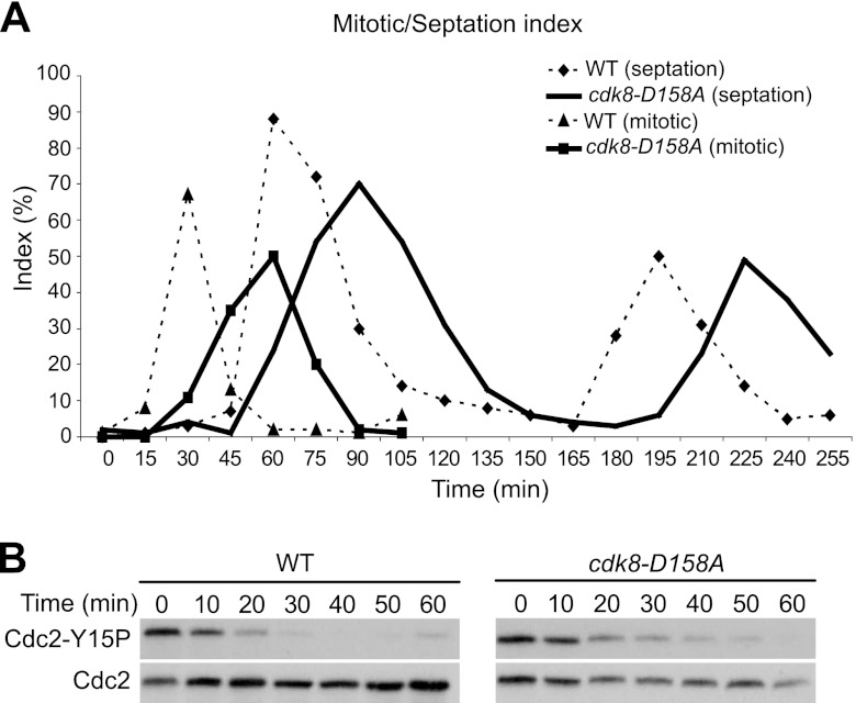
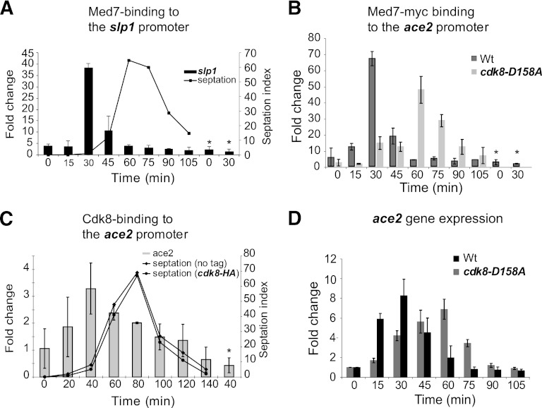
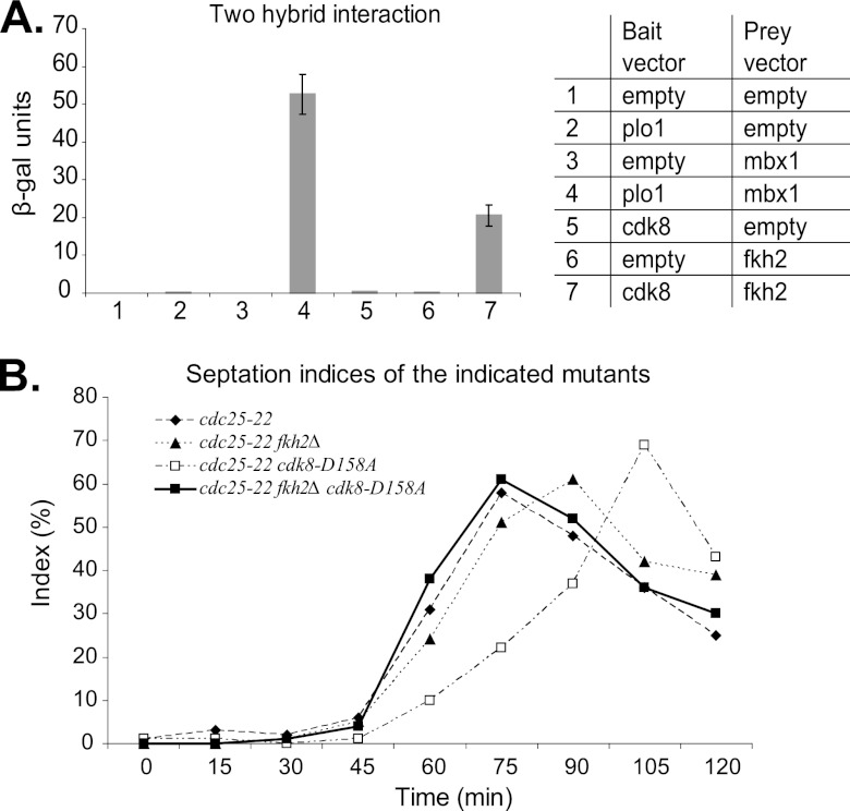
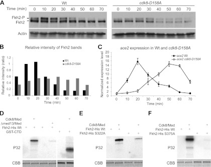
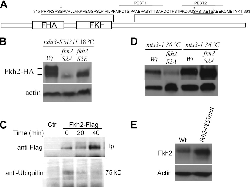
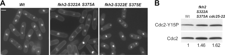
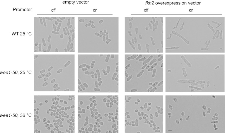
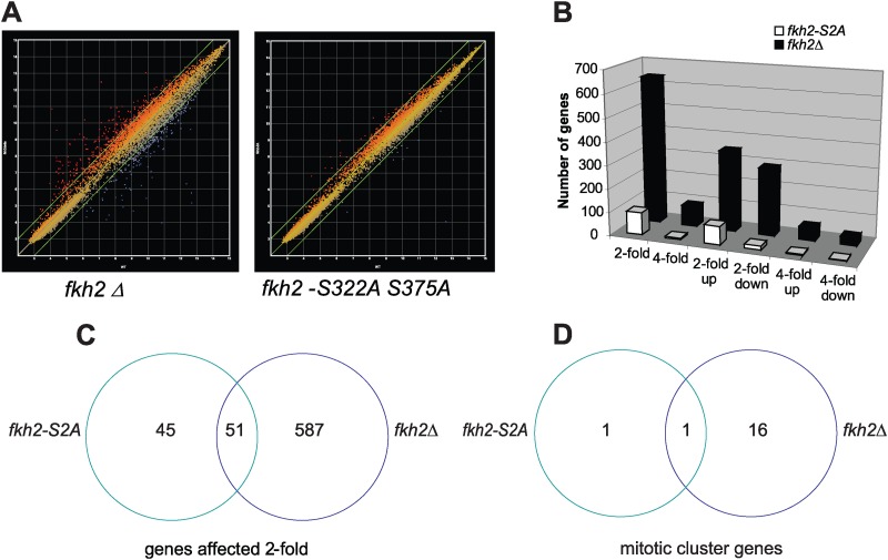
Temporal changes in transcription programs are coupled to control of cell growth and division. We here report that Mediator, a conserved coregulator of eukaryotic transcription, is part of a regulatory pathway that controls mitotic entry in fission yeast. The Mediator subunit cyclin-dependent kinase 8 (Cdk8) phosphorylates the forkhead 2 (Fkh2) protein in a periodic manner that coincides with gene activation during mitosis. Phosphorylation prevents degradation of the Fkh2 transcription factor by the proteasome, thus ensuring cell cycle-dependent variations in Fkh2 levels. Interestingly, Cdk8-dependent phosphorylation of Fkh2 controls mitotic entry, and mitotic entry is delayed by inactivation of the Cdk8 kinase activity or mutations replacing the phosphorylated serine residues of Fkh2. In addition, mutations in Fkh2, which mimic protein phosphorylation, lead to premature mitotic entry. Therefore, Fkh2 regulates not only the onset of mitotic transcription but also the correct timing of mitotic entry via effects on the Wee1 kinase. Our findings thus establish a new pathway linking the Mediator complex to control of mitotic transcription and regulation of mitotic entry in fission yeast.


Figures

Fig 1
Fig 1
Cdk8 regulates mitotic entry. (A) The mitotic and septation indices of synchronized wild-type and cdk8-D158A mutant cells. (B) Measurement of Cdk1-Y15 phosphorylation in synchronously dividing wild-type and cdk8-D158A cells.
Fig 2
Fig 2
Cdk8/Mediator binds to mitotic promoters in wt and cdk8-D158A mutant cells. Data represent the results of ChIP analysis performed using synchronously dividing cells. Samples were collected at the indicated time points. Asterisks indicate time points corresponding to an untagged control strain. (A) Med7-myc binding to the mitotic slp1 promoter in wt cells. (B) Med7-myc binding to the mitotic ace2 promoter in wt and cdk8-D158A cells. (C) Cdk8-HA binding to the ace2 promoter in wt cells. Septation indices are included to show that the HA tag does not affect cell cycle progression. (D) Real-time PCR measurement of ace2 transcription. Peaks of mRNA levels in both wt and cdk8-D158A cells coincided with the peak of Mediator complex binding. All measurements were repeated three times, and the mean values are shown, with error bars representing standard deviations.
Fig 3
Fig 3
Cdk8 physically and functionally interacts with transcription factor Fkh2. (A) Two-hybrid interaction between Cdk8 and Fkh2. β-Galactosidase (β-Gal) assays were performed in triplicate, and error bars represent standard deviations. The table displays the combinations of different plasmids used in two-hybrid analysis. As a positive control, we used Plo1 and Mbx1, which have previously been described as interacting (43). (B) The septation indices of the indicated mutants were followed in synchronized cell cultures. The delay of septation in the cdk8-D158A mutant was suppressed by the deletion of fkh2.
Fig 4
Fig 4
Cdk8 phosphorylates Fkh2 in vitro and in vivo. (A) Periodic phosphorylation of Fkh2 during mitosis in synchronized wt and cdk8-D158A mutant cells. Fkh2-HA and actin were revealed by immunoblotting. Please note that the protein extracts were separated on one single SDS-PAGE gel, followed by immunoblotting on one membrane, thus allowing direct comparison of protein levels between wt and cdk8-D158A mutant cells. (B) The relative intensities of the phosphorylated and nonphosphorylated Fkh2 bands displayed in panel A. (C) Transcription of the ace2 mitotic gene in wt and cdk8-D158A cells, measured by real-time PCR in the same synchrony experiment as described for panel A. (D) In vitro kinase assay performed with purified recombinant Fkh2. Mediator complexes containing Cdk8 (Cdk8/Med) or depleted of Cdk8 (Δmed13/Med) were incubated with recombinant Fkh2 in the presence of [γ-32P]ATP. The phosphorylated proteins were separated by SDS-polyacrylamide gel electrophoresis and identified by autoradiography (P32) or Coomassie staining (CBB). (E) In vitro kinase assay performed as described for panel B but with recombinant Fkh2 S322A. (F) In vitro kinase assay performed as described for panel B but with recombinant Fkh2 S375A.
Fig 5
Fig 5
Cdk8 phosphorylates Fkh2 in vivo and controls protein stability via the proteasome. (A) Schematic representation of the Fkh2 protein, displaying the forkhead-associated (FHA) and the forkhead (FKH) domains. The sequence view shows the phosphorylated residues (asterisks) and the PEST sequence motifs that were identified using the epestfind algorithm. The boxed sequence was deleted in the fkh2-PESTmut strain. (B) Immunoblot analysis of Fkh2 arrested in early mitosis performed using the cold-sensitive β-tubulin mutant nda3-KM311 mutation at 18°C. Antibodies against the HA tag were used to analyze levels of wt Fkh2, Fkh2-S2A, and Fkh2-S2E. Actin is visualized as a loading control. (C) Ubiquitylation of Fkh2 during mitosis. Fkh2-FLAG and untagged control (Ctr) cells were synchronized using cdc25-22 block release, and samples were collected for FLAG immunoprecipitation (Ip) and probed with anti-FLAG and antiubiquitylation antibodies to detect Fkh2 and the ubiquitin modification. (D) Western blot analysis of Fkh2 or Fkh2-S2A in mts3-1 mutant cells after incubation for 6 h at 30°C or 36°C. (E) Immunoblot analysis of Fkh2 protein levels in nonsynchronized wt and fkh2-PESTmut strains. Actin is shown as a loading control.
Fig 6
Fig 6
Fkh2 influences mitotic entry. (A) Microscopic images of wild-type, fkh2-S322A S375A, and fkh2-S322E S375E mutant cells. Nuclei and septa were stained. Delayed or advanced mitotic entry caused an increase or decrease of cell size at division in the mutants. Bar, 3.5 μm. (B) Western analysis of Cdc2-Y15 levels in the indicated strains, asynchronously dividing at 25°C. The relative intensities of Cdc2-Y15 compared to the total Cdc2 protein levels are indicated under the panel.
Fig 7
Fig 7
Overexpression of fkh2 in wt and wee1-50 cells. Cells were transformed with a vector containing the fkh2 gene under the control of the nmt1 promoter or empty vector as a control. Cells were propagated overnight under induced or noninduced conditions at 25°C or 36°C. Bar, 5 μm.
Fig 8
Fig 8
Microarray profile of fkh2Δ and fkh2-S322A S375A (S2A) mutants. (A) Scatter plot image of changes in gene expression. The two oblique lines represent a 2-fold cutoff. (B) Chart showing the number of genes affected in the fkh2Δ and fkh2-S2A mutants. (C and D) Venn diagrams showing total numbers representing overlap of the genes (C) and overlap of mitotic cluster genes (D) that changed more than 2-fold in the fkh2Δ and fkh2-S2A mutants.
Fig 9
Fig 9
Fkh2 phosphorylation status affects promoter binding. (A) ChIP analysis of Fkh2 binding to the ace2 promoter during mitosis in synchronized wt and cdk8-D158A mutant cells. Samples were collected at the indicated time points. (B) ChIP analysis of Fkh2, Fkh2-S2A, and Fkh2-S2E binding to the ace2 promoter. Cells were either nonsynchronized (NS) or arrested in early mitosis (M) by the use of the cold-sensitive β-tubulin mutant nda3-KM311 mutation at 18°C.

Similar articles

Cited by

References

    1. Agarwal M, et al. 2010. Mid1p-dependent regulation of the M-G1 transcription wave in fission yeast. J. Cell Sci. 123:4366–4373 - PubMed
    1. Alarcón C, et al. 2009. Nuclear CDKs drive Smad transcriptional activation and turnover in BMP and TGF-beta pathways. Cell 139:757–769 - PMC - PubMed
    1. Almonacid M, Paoletti A. 2010. Mechanisms controlling division-plane positioning. Semin. Cell Dev. Biol. 21:874–880 - PubMed
    1. Anderson M, et al. 2002. Plo1(+) regulates gene transcription at the M-G(1) interval during the fission yeast mitotic cell cycle. EMBO J. 21:5745–5755 - PMC - PubMed
    1. Bähler J. 2005. Cell-cycle control of gene expression in budding and fission yeast. Annu. Rev. Genet. 39:69–94 - PubMed

Publication types

MeSH terms

Substances