. 2012 Jun 8;287(24):20636-51.
doi: 10.1074/jbc.M111.314997. Epub 2012 Mar 26.

Engulfment protein GULP is regulator of transforming growth factor-β response in ovarian cells

Affiliations

Engulfment protein GULP is regulator of transforming growth factor-β response in ovarian cells

Cheng-I J Ma et al. J Biol Chem. .

Abstract

Transforming growth factor β (TGF-β) is a key regulatory molecule with pleiotropic effects on cell growth, migration, and invasion. As a result, impairment of proper TGF-β signaling is central to tumorigenesis and metastasis. The TGF-β receptor V (TGFBRV or LRP1) has been shown to be responsible for TGF-β-mediated cell growth inhibition in Chinese hamster ovary (CHO) cells. The LRP1 adapter protein GULP mediates internalization of the various LRP1-specific ligands, and we hypothesize that GULP acts as a novel regulator of TGF-β signaling in ovarian cells. CHO cells that overexpress exogenous GULP (FL) demonstrate enhancement in growth inhibition, migration, and invasion from TGF-β treatment, whereas cells that lack GULP (AS) show impairment of growth inhibition and decreased migration and invasion. The enhanced TGF-β response in FL cells was confirmed by a prolonged TGF-β-induced SMAD3 phosphorylation, whereas a shortening of the phosphorylation event is observed in AS cells. Mechanistically, the presence of GULP retains the TGF-β in a signaling-competent early endosome for enhanced signaling. To address this mechanism in a physiological setting, TGF-β insensitive ovarian adenocarcinoma cells (HEY) have a very low GULP expression level, similar to the observation made in a wide selection of human ovarian adenocarcinomas. Transfection of GULP into the HEY cells restored the TGF-β responsiveness, as measured by SMAD3 phosphorylation and impairment of cell growth. Because GULP expression positively regulates TGF-β signaling leading to growth inhibition, this may represent an attractive target to achieve TGF-β responsiveness in ovarian cells.


Figures

FIGURE 1.
FIGURE 1.
The growth-inhibitory effect of TGF-β in CHO cells correlates to the expression level of GULP. A, control CHO cells (WT), CHO cells overexpressing GULP (FL), CHO cells with reduced GULP expression (AS), and LRP1-deficient CHO cells (13-5-1) were treated with TGF-β or not for 48 h. Cells were resuspended and counted using a hemacytometer. Values are expressed as the percentage decrease of the number of cells between the conditions with and without TGF-β. Results presented here are the mean ± S.D. (error bars) of three separate experiments. *, p < 0.003; **, p < 0.0001 in comparison with WT. B, WT, FL, AS, and 13-5-1 cells were stimulated without or with TGF-β for 72 h before cell growth was measured by a cell viability assay (MTT). Values were computed and presented as percentage of growth inhibition induced by TGF-β as described under “Materials and Methods.” Results shown here are the mean ± S.D. of two separate experiments, each performed in quadruplicate. *, p < 0.03; **, p < 0.01; ***, p < 0.002 in comparison with WT. C, WT, FL, AS, and 13-5-1 cells were stimulated without or with TGF-β for 24 h, and then an apoptosis kit assay (Sigma) was performed according to manufacturer's instructions. Results are presented as the mean percentage of total cells ± S.D. that stain positive for Annexin V of five separate plates of at least 200 cells/plate for each condition. D, control MEF cells (MEF-1), MEF cells with a genetic deficiency of GULP (GULP KO), GULP KO cells with reconstituted GULP expression (reconst. GULP), and LRP1-deficient MEF cells (MEF-2) were treated without or with TGF-β for 48 h, and then cells were counted as in A. *, p < 0.01 comparing TGF-β-treated and -untreated conditions. E, FL or AS cells were treated without or with TGF-β for 16 h; cell lysates were harvested; and Western blots of p21Cip1, p15ink4b, and tubulin were performed.
FIGURE 2.
FIGURE 2.
Overexpression of GULP in CHO cells leads to enhanced cell motility and invasion in response to TGF-β treatment. A, transwell migration assays were conducted using WT, FL, and AS cells. Results show migration (as measured by crystal violet color intensity) for TGF-β-untreated (Control) and -treated cells (TGF-β). Data presented here are the mean ± S.D. (error bars) of three experiments, each done in triplicate. *, p < 0.003; **, p < 0.0003 in comparison with the untreated condition. B, wound healing assays were conducted using WT, FL, and AS cells. Cells were grown to confluence and serum-starved for 24 h, and then a scratch of ∼1 mm was introduced through the middle of the plate. Cells were then treated without or with TGF-β for 24 h, and the percentage of area that TGF-β-induced cells grow into the scratch (wound healing) was measured and computed as described under “Materials and Methods.” Results presented here are the mean ± S.D. of three experiments, each done in triplicate. *, p ≤ 0.001 in comparison with the untreated condition. C, Transwell invasion assays were conducted using WT, FL, and AS cells without and with TGF-β treatment for 24 h. Cell invasion (the number of cells that crossed the Matrigel barrier) was measured by the intensity of crystal violet staining in the well. Data presented here were the mean ± S.D. of two experiments, each done in triplicate. *, p ≤ 0.004 in comparison with the untreated condition. D, gelatin zymography experiments were performed on media from WT, FL, and AS cells treated without or with TGF-β for 24 h. To quantify the extent of active MMP-9 secreted, densitometry was performed on the MMP-9 digestion band in the absence and presence of TGF-β and presented as the mean ± S.D. of three separate gelatin zymography experiments. *, p < 0.03; ***, p < 0.0003.
FIGURE 3.
FIGURE 3.
GULP signaling involves TGF-β·RI. A, WT and FL cells were transfected with scrambled RNA or siRNA specific for TGF-β·RI for 16 h and then were treated without or with TGF-β for 60 min. Total cell lysates were collected and analyzed by Western blot using anti- TGF-β·RI, anti-pSMAD3, and anti-SMAD antibodies. TGF-β·RI expression is efficiently prevented by siRNA, which also prevents pSMAD3 formation. B, a growth inhibition experiment (similar to Fig. 1) was performed. WT and FL cells were transfected with scrambled RNA or siRNA specific for TGF-β·RI for 16 h and then treated without or with TGF-β for 24 h. Cells were resuspended and counted using a hemacytometer. Values are expressed as the percentage decrease of the number of cells between the conditions with and without TGF-β. Results presented here are the mean ± S.D. (error bars) of three separate experiments.
FIGURE 4.
FIGURE 4.
Enhanced GULP expression results in prolonged SMAD3 phosphorylation and increased TGF-β activity. A, WT, FL, AS, and 13-5-1 cells were treated with TGF-β for the time periods indicated. Cell lysates were analyzed by Western blot using anti-pSMAD3 antibody (Cell Signaling), SMAD3 antibody (Cell Signaling), and GAPDH antibody (Santa Cruz Biotechnology, Inc.). Positive controls were included for AS and 13-5-1 cells to prove that the Western blots were detecting properly. B, WT, FL, AS, and 13-5-1 cells were stimulated with TGF-β for 240 min, and the cell lysates were analyzed by Western blot using anti-phospho-SMAD3 antibody, SMAD3 antibody, and GAPDH antibody. A representative blot is shown at the top, including a positive control. Densitometric scaling was performed to normalize the intensity of pSMAD3 to SMAD3 expression. The results were plotted as relative intensity of pSMAD3, with the intensity of WT being normalized to 1, and three independent Western blot analyses generated the mean ± S.D. (error bars). *, p ≤ 0.05. C, WT, FL, AS, and 13-5-1 cells were co-transfected with a 3TP-Lux reporter construct and a β-galactosidase expression plasmid. Cells were treated with TGF-β or not for 16 h, and luciferase assays were performed. Data were presented as -fold increase in activity of TGF-β-treated samples compared with the non-treated WT control. The experiments were repeated three times, each done in triplicate, and results presented here are the mean ± S.D. from three experiments. In comparison with the -fold increase in TGF-β activity in WT, significance is shown. *, p < 0.05; ***, p < 0.001. D, FL and AS cells were stimulated without or with TGF-β for 60 min, and the cell lysates were analyzed by Western blot using anti-pSMAD2 antibody, SMAD2 antibody, and tubulin antibody. The experiment was repeated three times, and a representative blot is shown.
FIGURE 5.
FIGURE 5.
GULP expression is inducible by TGF-β. A (top), WT cells were stimulated with TGF-β for a time course as indicated, and total cell lysates were analyzed by Western blot using anti-GULP, anti-Dab2, and anti-GAPDH antibodies. This experiment was performed three times, and a representative Western blot is shown here. A (bottom), to quantify the bands using densitometry, we measured the relative intensity of the GULP band after normalization to the intensity of GAPDH and then calculated the average ± S.D. (error bars). *, p < 0.05; **, p < 0.005; ***, p < 0.001. B, WT cells were stimulated with TGF-β for 24 h, and then mRNAs were extracted, and reverse transcription PCR was performed. Resulting cDNAs were then amplified using the primers specific for GULP, Dab2, and GAPDH for 25 cycles.
FIGURE 6.
FIGURE 6.
Ovarian adenocarcinoma cells have a low expression level of GULP. A, microarray data on the relative expression level of GULP from normal ovary and several different ovarian adenocarcinomas was analyzed and plotted into box plots (number of samples is shown below the bar; original data are from Schwartz et al. (43), with permission from authors). The values are displayed at a base 2 logarithmic transformation. B, SKOV3 and HEY cells were characterized in terms of TGF-β sensitivity. Total cell lysates of SKOV3 and HEY were collected after 45 min of TGF-β treatment and were analyzed by Western blot using anti-pSMAD3, SMAD3, and anti-GAPDH antibodies. C, SKOV3 and HEY cells were stimulated with TGF-β for 0 and 8 h. Total cell lysates were analyzed by Western blot using anti-GULP and anti-GAPDH antibodies. Sample blots are displayed here. The experiment was replicated three times, and densitometry was used to measure the band intensities (normalized to GAPDH), to generate the average intensity ± S.D. (error bars).
FIGURE 7.
FIGURE 7.
Enhanced GULP expression in TGF-β-unresponsive HEY cells results in increased TGF-β activity. A, HEY cells, HEY cells transiently expressing YFP (HEY-YFP), and HEY cells transiently expressing YFP-GULP (HEY-GULP) were stimulated with TGF-β for 120 min. Total cell lysates were collected and analyzed by Western blot using anti-pSMAD3, anti-SMAD3, anti-GAPDH, and anti-YFP antibodies. Densitometric scaling was performed to normalize the intensity of pSMAD3 to total SMAD3 levels. The results are plotted below as relative intensity of pSMAD3 normalized to untransfected HEY cells. Data presented here are the mean ± S.D. (error bars) of three independent analyses, with a representative blot shown at the top. *, p < 0.03. B, HEY cells were transfected with a YFP expression plasmid or transfected with a plasmid expressing YFP-GULP. Transfected cells were treated without or with TGF-β for 48 h, and cell growth was measured by the cell viability MTT assay. Values were computed and presented as percentage of growth inhibition induced by TGF-β as described under “Materials and Methods.” Results shown here are the mean ± S.D. of two independent experiments, each performed with six replicates. *, p < 0.0001. C, SKOV3 cells were transfected with scrambled RNA (scRNA) or siRNA to GULP for 24 h, and then they were treated with TGF-β for 60 min. Cell lysates were prepared, and Western blots of pSMAD3, SMAD3, and GULP were performed. Relative intensities of pSMAD3 as measured by densitometry (normalized to total SMAD3 levels) were measured, and the average ± S.D. is presented here. **, p < 0.01 in comparison with the no treatment condition; ***, p < 0.002 between the scrambled RNA and siRNA conditions. D, SKOV3 cells were transfected with GULP siRNA or a scrambled control (scRNA). Transfected cells were treated without or with TGF-β for 48 h, and cell growth was measured by the cell viability MTT assay. Values were computed and presented as percentage of growth inhibition induced by TGF-β as described under “Materials and Methods.” Results shown here are the mean ± S.D. of two independent experiments, each performed with six replicates. *, p < 0.05.
FIGURE 8.
FIGURE 8.
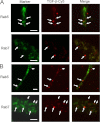
TGF-β is trapped in the early endosome in FL cells. FL cells (A) or AS cells (B) were transfected with Rab5 (early endosome marker) or Rab7 (late endosome marker) to label endosome compartments. These cells were incubated with Cy5-labeled TGF-β (red fluorescence) for 1 h and then analyzed by fluorescence microscopy. Overlap of TGF-β in an endosome compartment can be determined by examining the white arrow in the separate frames (as an example). FL cells show considerable overlap of Rab5 endosomes and TGF-β but no overlap of Rab7 endosomes and TGF-β. AS cells show little overlap of Rab5 endosomes and TGF-β but significant overlap of Rab7 endosomes and TGF-β. The white arrows indicate endosomal TGF-β. White bar, 10 μm.
FIGURE 9.
FIGURE 9.
GULP affects trafficking and degradation of LRP ligands. A, WT-, FL-, or PTB (the dominant negative mutant that behaves similarly to AS cells)-expressing cells were incubated with 125I-labeled methylamine-activated α2M at 37 °C for various times. At each time point, cell medium was collected, and TCA-soluble 125I was detected (corresponding to degraded ligand). The most radioactivity was observed in PTB-expressing cells, demonstrating that LRP ligands are internalized and preferentially delivered to the lysosome in PTB-expressing cells. B, FL- and AS-expressing cells were incubated with 125I-labeled TGF-β at 37 °C for various times in the absence or presence of competing amounts of RAP. At each time point, cell medium was collected, and TCA-soluble 125I was detected. AS cells had the most radioactivity, indicating the most degradation of ligand, which could be effectively competed by the addition of RAP. C and D, PEA is an LRP ligand that, after internalization and trafficking to the late endosome, escapes the late endosome and becomes cytotoxic. WT, FL, AS, and 13-5-1 cells were incubated without (PEA Null) or with PEA (PEA), for 24 h and then cells were counted. C, actual cell counts normalized to WT at 100 cells. D, ratio of PEA/PEA null for each cell type. FL cells are only partially affected by PEA, whereas AS cells are extremely sensitive to PEA. *, p < 0.01. Error bars, S.D.
FIGURE 10.
FIGURE 10.
Schematic model of the role of GULP in the trafficking of LRP ligands. Degradation of LRP ligands requires the timely and coordinated recruitment of adapter and effector proteins to mediate internalization and trafficking of the LRP ligands to the lysosome (top). Dab2 is positioned at the plasma membrane to recruit LRP to clathrin-coated pits. AP-2 binds clathrin and mediates inward curvature to generate an endosome. At the early endosome, GULP binds LRP and recruits ARF6 (GDP-bound) and ACAP1 (GAP specific to ARF6), thereby excluding ARNO (guanine nucleotide exchange factor specific to ARF6), vacuolar H+-ATPase, and the PI 5-kinase (PI-5-K). Exclusion of ARNO and PI 5-kinase prevents maturation of the early endosome to a late endosome (bottom) and prevents efficient sorting of the receptor (to be recycled) and ligand (to be degraded). Thus, in the presence of GULP, TGF-β is trapped in the early endosome and consequently promotes enhanced TGF-β signaling.

Similar articles

Cited by

References

    1. Roberts A. B., Sporn M. B. (1985) Transforming growth factors. Cancer Surv. 4, 683–705 - PubMed
    1. Sporn M. B., Roberts A. B., Wakefield L. M., Assoian R. K. (1986) Transforming growth factor-β. Biological function and chemical structure. Science 233, 532–534 - PubMed
    1. Zhou L., Leung B. S. (1992) Growth regulation of ovarian cancer cells by epidermal growth factor and transforming growth factors α and β1. Biochim. Biophys. Acta 1180, 130–136 - PubMed
    1. Massagué J. (1998) TGF-β signal transduction. Annu. Rev. Biochem. 67, 753–791 - PubMed
    1. Blobe G. C., Schiemann W. P., Lodish H. F. (2000) Role of transforming growth factor β in human disease. N. Engl. J. Med. 342, 1350–1358 - PubMed

MeSH terms