. 2012 May 18;287(21):17471-17482.
doi: 10.1074/jbc.M112.352781. Epub 2012 Mar 27.

Cathepsins L and Z are critical in degrading polyglutamine-containing proteins within lysosomes

Affiliations

Cathepsins L and Z are critical in degrading polyglutamine-containing proteins within lysosomes

Nidhi Bhutani et al. J Biol Chem. .

Abstract

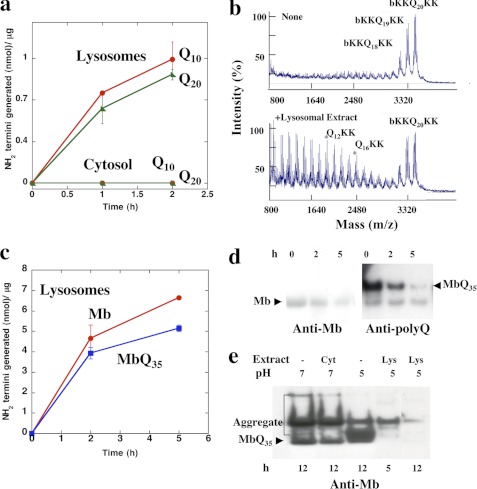
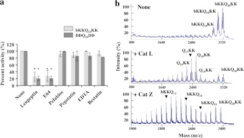
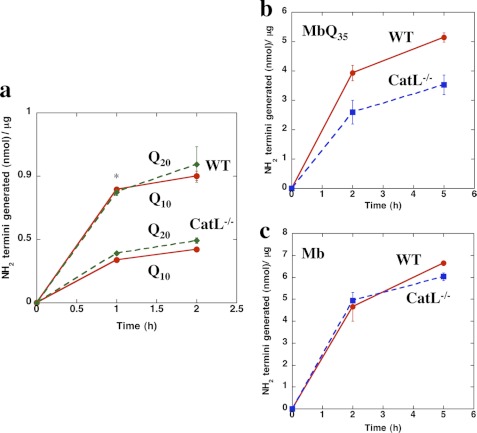
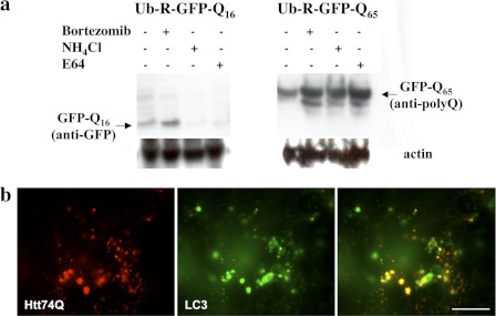
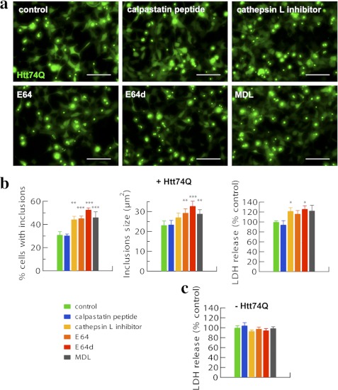
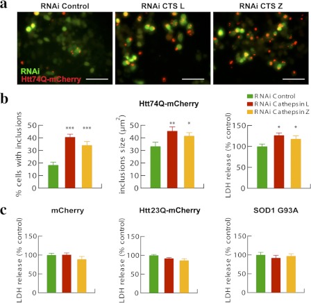
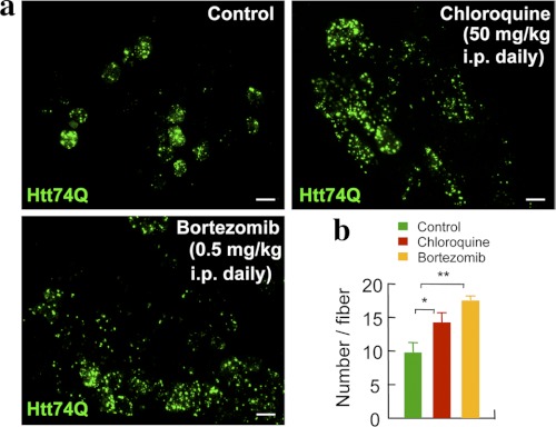
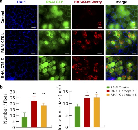
In neurodegenerative diseases caused by extended polyglutamine (polyQ) sequences in proteins, aggregation-prone polyQ proteins accumulate in intraneuronal inclusions. PolyQ proteins can be degraded by lysosomes or proteasomes. Proteasomes are unable to hydrolyze polyQ repeat sequences, and during breakdown of polyQ proteins, they release polyQ repeat fragments for degradation by other cellular enzymes. This study was undertaken to identify the responsible proteases. Lysosomal extracts (unlike cytosolic enzymes) were found to rapidly hydrolyze polyQ sequences in peptides, proteins, or insoluble aggregates. Using specific inhibitors against lysosomal proteases, enzyme-deficient extracts, and pure cathepsins, we identified cathepsins L and Z as the lysosomal cysteine proteases that digest polyQ proteins and peptides. RNAi for cathepsins L and Z in different cell lines and adult mouse muscles confirmed that they are critical in degrading polyQ proteins (expanded huntingtin exon 1) but not other types of aggregation-prone proteins (e.g. mutant SOD1). Therefore, the activities of these two lysosomal cysteine proteases are important in host defense against toxic accumulation of polyQ proteins.


Figures

FIGURE 1.
FIGURE 1.
Unlike cytosolic extracts, lysosomes rapidly digest polyQ peptides and proteins. a, b-KKQ10KK or b-KKQ20KK (15 μm) was incubated with either 3.2 μg of cytosolic extracts in a 30-μl reaction in 50 mm sodium phosphate, 1 mm DTT, pH 7, or with 3.2 μg of lysosomal extracts in a 30-μl reaction in 50 mm sodium acetate, 1 mm DTT, pH 5, at 37 °C, and the new N termini generated were assayed with fluorescamine. b, b-KKQ20KK (15 μm) was incubated alone or with 3.2 μg of lysosomal extracts in 50 mm sodium acetate, 1 mm DTT, pH 5, at 37 °C for 2 h, and the reaction mixtures were analyzed by MALDI-TOF. b-KKQ20KK alone (same as time (t) = 0 for the reaction) contains contaminating peptides b-KKQ19KK and b-KKQ18KK. c, WT Mb or Mb-Q35 fusion protein (MbQ35) (20 μm) was incubated with 3.2 μg of lysosomal extracts in a 30-μl reaction in 50 mm sodium acetate, 1 mm DTT, pH 5, at 37 °C, and the new N termini generated were assayed with fluorescamine. d, 20 μm WT Mb and Mb-Q35 were incubated with lysosomal extracts, and the reaction aliquots were boiled in 1× SDS sample buffer containing 100 mm DTT, separated by SDS-PAGE, and probed with either a polyclonal antibody against myoglobin or a monoclonal antibody against the polyQ sequence. For Mb-Q35, only the anti-polyQ blot is shown for simplicity. e, Mb-Q35 was preaggregated in 50 mm sodium phosphate, 1 mm DTT, pH 7, for 8 h at 37 °C and then either incubated alone at pH 7 or pH 5 or with 3.2 μg of cytosolic extracts at pH 7 or 3.2 μg of lysosomal extracts in 50 mm sodium acetate, 1 mm DTT, pH 5, at 37 °C. The reaction products were analyzed at the indicated times by SDS-PAGE followed by immunoblotting with anti-myoglobin antibodies. The labeled band corresponds to the monomeric Mb-Q35. Error bars, S.D.
FIGURE 2.
FIGURE 2.
Lysosomal cysteine proteases are responsible for the digestion of polyQ peptides. a, b-KKQ10KK and DDQ20DD (15 μm) were incubated with 3.2 μg of lysosomal extracts in 50 mm sodium acetate, 1 mm DTT, pH 5, at 37 °C for 1 h, in the presence or absence of inhibitors, and the new N termini generated were assayed. The lysosomal extracts were preincubated with the inhibitors for 20 min at room temperature. The concentrations used were 100 μm for leupeptin, 100 μm for E64, 1 mm for pefabloc, 100 μm for pepstatin, 1 mm for EDTA, and 100 μm for bestatin. *, p < 0.001. b, b-KKQ20KK (15 μm) was incubated with activated pure recombinant human cathepsins B, L, H, S, and Z at amounts varying between 10 and 100 ng under optimal reaction conditions for each cathepsin at 37 °C, and the reaction mixtures were analyzed by MALDI-TOF. Digestion patterns for b-KKQ20KK alone (same as t = 0 of the reaction) and with cathepsin L and cathepsin Z are shown. No digestion of b-KKQ20KK was observed by cathepsins B, S, and H. Cathepsin L acts as an endopeptidase in digesting b-KKQ20KK, whereas cathepsin Z digests b-KKQ20KK as a carboxypeptidase. Error bars, S.D.
FIGURE 3.
FIGURE 3.
Lysosomal extracts from Cathepsin L−/− mice cannot efficiently degrade long-polyQ peptides and proteins. a, b-KKQ10KK and b-KKQ20KK (15 μm) were incubated with 3.2 μg of lysosomal extracts prepared from WT mice or cathepsin L−/− mice in 50 mm sodium acetate, 1 mm DTT, pH 5, at 37 °C, and the new N termini generated were assayed. *, p < 0.01. b and c, WT-Mb (c) or Mb-Q35 (b) (20 μm) was incubated with 3.2 μg of lysosomal extracts prepared from WT mice or cathepsin L−/− mice in 50 mm sodium acetate, 1 mm DTT, pH 5, at 37 °C, and the new N termini generated were assayed. Error bars, S.D.
FIGURE 4.
FIGURE 4.
Short polyQ proteins are degraded by proteasomes, whereas both proteasomes and lysosomal cysteine proteases degrade long polyQ proteins in vivo. a, HeLa cells transiently transfected for 36 h with Ub-R-GFPQ16 and Ub-R-GFPQ65 were treated with specific inhibitors for 12 h (i.e. 10 μm bortezomib, 15 mm NH4Cl, and 100 μm E64). Soluble cell extracts following a 10,000 × g centrifugation were analyzed by immunoblotting with antibodies against GFP or polyQ sequences. Anti-actin blots showed similar intensities for all of the lanes. b, NIH3T3 cells were transfected with Htt74Q-mCherry and CFP-LC3 to stain lysosomes. For simplicity, lysosomes are shown in green, so that areas of co-localization appear in yellow. Scale bar, 20 μm.
FIGURE 5.
FIGURE 5.
Cathepsin (cysteine protease) inhibitors but not calpain inhibitors increase the proportion of cells with inclusions, the sizes of inclusions, and the toxicity in HEK293A cells expressing expanded huntingtin exon 1. a, HEK293A cells expressing Htt74Q-GFP for 48–72 h were treated with the indicated inhibitors for 24 h (see “Experimental Procedures”). Representative pictures are shown. Scale bar, 100 μm. b, quantitation of the number of inclusions displayed by HEK293A cells expressing Htt74Q-GFP following different treatments is reported as well as that for sizes of inclusions and related cell toxicity in lactate dehydrogenase (LDH) release assays. Error bars, S.D. *, p < 0.05; **, p < 0.01; ***, p < 0.001. n = 6. c, HEK293A cells not expressing Htt74Q-GFP were treated as above, and cell toxicity was measured in lactate dehydrogenase release assays. No evident cell toxicity per se was observed with any of the inhibitors used. n = 6.
FIGURE 6.
FIGURE 6.
Reduced expression of cathepsin L or Z increases the proportion of cells with inclusions, the sizes of inclusions, and toxicity in neuroblastoma N1-E115 cells expressing expanded huntingtin exon 1. a, N1-E115 cells were transfected for 48 h with Htt74Q-mCherry along with GFP-RNAi constructs, either control or against cathepsin L or Z. Representative pictures are shown. Scale bar, 100 μm. b, quantitation of the number of inclusions displayed by N1-E115 cells expressing Htt74Q-mCherry following different treatments is reported as well as that for sizes of inclusions and related cell toxicity in lactate dehydrogenase (LDH) release assays. Error bars, S.D. *, p < 0.05; **, p < 0.01; ***, p < 0.001. n = 6. c, N1-E115 cells expressing mCherry, Htt23Q-mCherry, or SOD1G93A were tested for cell toxicity through lactate dehydrogenase release assays. No evident toxicity was observed for any of the proteins expressed. Error bars, S.D. n = 6.
FIGURE 7.
FIGURE 7.
In vivo inhibition of the lysosomal or the proteasomal pathway results in increased number of inclusions in mouse muscle fibers expressing expanded huntingtin exon 1. a, mice electroporated with plasmids for Htt74Q-GFP into the tibialis anterior muscle were injected with chloroquine or bortezomib daily for 7 days at the indicated doses. A representative picture for each treatment is shown. Scale bar, 50 μm. b, quantitation of the number of Htt74Q-GFP-positive inclusions per muscle fiber is reported for the different treatments. Error bars, S.D. *, p < 0.05; **, p < 0.01. n = 6.
FIGURE 8.
FIGURE 8.
Reduced expression of cathepsin L or Z increases the number and size of inclusions in mouse muscle fibers expressing expanded huntingtin exon 1. a, tibialis anterior muscles were electroporated for 7 days with plasmids expressing Htt74Q-mCherry along with GFP-RNAi constructs, either control or against cathepsin L or Z. A representative field of a transverse section of fibers for each condition tested is reported. Nuclei were stained with DAPI. Scale bar, 100 μm. b, quantitation of the number of inclusions per muscle fiber expressing Htt74Q-mCherry under the different treatments is reported as well as that for sizes of inclusions. Larger and more abundant aggregates are observed when cathepsin L or Z is depleted. Error bars, S.D. *, p < 0.05; **, p < 0.01. n = 6.

References

    1. Zoghbi H. Y., Orr H. T. (2000) Glutamine repeats and neurodegeneration. Annu. Rev. Neurosci. 23, 217–247 - PubMed
    1. Rubinsztein D. C. (2006) The roles of intracellular protein-degradation pathways in neurodegeneration. Nature 443, 780–786 - PubMed
    1. Kitamura A., Kubota H. (2010) Amyloid oligomers. Dynamics and toxicity in the cytosol and nucleus. FEBS J. 277, 1369–1379 - PubMed
    1. Davies S. W., Turmaine M., Cozens B. A., DiFiglia M., Sharp A. H., Ross C. A., Scherzinger E., Wanker E. E., Mangiarini L., Bates G. P. (1997) Formation of neuronal intranuclear inclusions underlies the neurological dysfunction in mice transgenic for the HD mutation. Cell 90, 537–548 - PubMed
    1. Waelter S., Boeddrich A., Lurz R., Scherzinger E., Lueder G., Lehrach H., Wanker E. E. (2001) Accumulation of mutant huntingtin fragments in aggresome-like inclusion bodies as a result of insufficient protein degradation. Mol. Biol. Cell 12, 1393–1407 - PMC - PubMed

Publication types

MeSH terms