. 2012 May 11;287(20):16709-19.
doi: 10.1074/jbc.M111.333781. Epub 2012 Mar 26.

1,3-1,4-α-L-fucosynthase that specifically introduces Lewis a/x antigens into type-1/2 chains

Affiliations

1,3-1,4-α-L-fucosynthase that specifically introduces Lewis a/x antigens into type-1/2 chains

Haruko Sakurama et al. J Biol Chem. .

Abstract

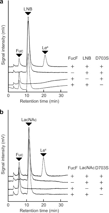
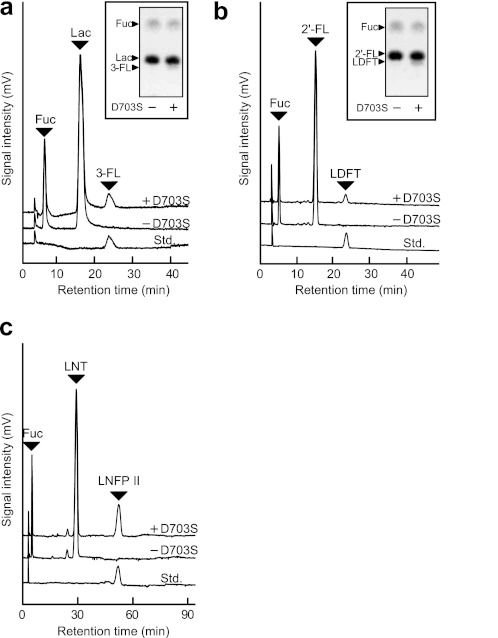
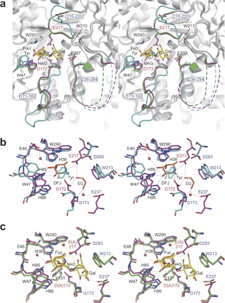
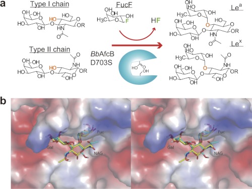
α-L-fucosyl residues attached at the non-reducing ends of glycoconjugates constitute histo-blood group antigens Lewis (Le) and ABO and play fundamental roles in various biological processes. Therefore, establishing a method for synthesizing the antigens is important for functional glycomics studies. However, regiospecific synthesis of glycosyl linkages, especially α-L-fucosyl linkages, is quite difficult to control both by chemists and enzymologists. Here, we generated an α-L-fucosynthase that specifically introduces Le(a) and Le(x) antigens into the type-1 and type-2 chains, respectively; i.e. the enzyme specifically accepts the disaccharide structures (Galβ1-3/4GlcNAc) at the non-reducing ends and attaches a Fuc residue via an α-(1,4/3)-linkage to the GlcNAc. X-ray crystallographic studies revealed the structural basis of this strict regio- and acceptor specificity, which includes the induced fit movement of the catalytically important residues, and the difference between the active site structures of 1,3-1,4-α-L-fucosidase (EC 3.2.1.111) and α-L-fucosidase (EC 3.2.1.51) in glycoside hydrolase family 29. The glycosynthase developed in this study should serve as a potentially powerful tool to specifically introduce the Le(a/x) epitopes onto labile glycoconjugates including glycoproteins. Mining glycosidases with strict specificity may represent the most efficient route to the specific synthesis of glycosidic bonds.


Figures

FIGURE 1.
FIGURE 1.
Specific syntheses of Lea (a) and Lex (b) trisaccharides by the BbAfcB D703S mutant. a, the reaction was carried out for 40 min at 30 °C in 100 mm MES buffer (pH 5.0) containing 40 mm FucF (donor), 200 mm LNB (acceptor), and a 17 μm concentration of the BbAfcB D703S mutant. For the control experiments, the substrate or enzyme was omitted from the reaction. b, for the synthesis of Lex, 100 mm LacNAc was used as the acceptor. The reaction products were analyzed by HPLC-CAD. The peaks of l-fucose (Fuc), LNB, LacNAc, Lea, and Lex are shown. Note that FucF is decomposed when terminating the reaction by trichloroacetic acid.
FIGURE 2.
FIGURE 2.
Acceptor specificity of BbAfcB D703S glycosynthase. The reaction was carried out in 100 mm MES buffer (pH 5.0) containing 40 mm FucF and a 100 mm concentration of each acceptor for 40 min at 30 °C in the presence and absence of the D703S mutant (17 μm). The acceptors used were as follows: Lac (a), 2′-FL (b), and LNT (c). The reaction products were analyzed by HPLC-CAD. The peaks of l-fucose (Fuc), acceptor, and standard sugars (Std.) are indicated. Inset in a and b are the results of the thin-layer chromatography (TLC) analysis of the reaction products. See also Table 1 and supplemental Figs. S3–S7.
FIGURE 3.
FIGURE 3.
Induced fit movement of BiAfcB upon substrate binding (stereoviews). a, superimposed structures of the ligand-free form (cyan; Protein Data Bank code 3MO4) (37), WT-DFJ-EG complex (protein in magenta; ligand in orange), and D172A/E217A-LNFP II complex (protein in green; ligand in yellow). The ligand-free structure contains a tyrosine molecule at the Fuc-binding site (subsite −1). The side chain of Trp-47 and two loop regions (173–182 and 215–220) are significantly displaced. Disordered regions of the 236–254 loop are shown as dotted lines. b and c, the structure of the substrate-binding site. b, ligand-free (cyan) and WT-DFJ-EG complex (protein in magenta; ligand in orange). The tyrosine molecule bound to the ligand-free structure is shown transparently. Water molecules and hydrogen bonds in the WT-DFJ-EG complex are shown. c, WT-DFJ-EG complex (protein in magenta; ligand in orange) and D172A/E217A-LNFP II complex (protein in green; ligand in yellow). Water molecules and hydrogen bonds in the LNFP II complex are shown. Ethylene glycol and GlcNAc are labeled as EG and NAG, respectively.
FIGURE 4.
FIGURE 4.
a, regio- and stereospecific installation of a Fuc residue into type-1/2 chains catalyzed by the BbAfcB D703S glycosynthase. b, the predicted mode of binding of Lex trisaccharide (cyan) and its comparison with the Lea trisaccharide (yellow) observed in the D172A/E217A-LNFP II structure (electrostatic surface potential map) (stereoview). NAG, GlcNAc.
FIGURE 5.
FIGURE 5.
Phylogenetic and structural difference between two subfamilies of GH29. a, the phylogenetic analysis of enzymatically characterized GH29 α-l-fucosidases. The tree was constructed using the ClustalW program with a neighbor-joining method (55). b, comparison of the catalytic sites of BiAfcB (D172A/E217A-LNFP II complex; protein in green; ligand in yellow) and TmFuc (Fuc complex; magenta; Protein Data Bank code 1ODU) (stereoview) (44). The numbers of the residues of BiAfcB and TmFuc are labeled in green and magenta, respectively. See also supplemental Figs. S11 and S12. A., Arabidopsis; H., Homo; R., Rattus; D. discoideum, Dictyostelium discoideum; C., Canis; L., Lactobacillus; B. infantis, Bifidobacterium longum subsp. infantis; B. thetaiotaomicron, Bacteroides thetaiotaomicron; D. melanogaster, Drosophila melanogaster.

References

    1. Becker D. J., Lowe J. B. (2003) Fucose: biosynthesis and biological function in mammals. Glycobiology 13, 41R–53R - PubMed
    1. Gooi H. C., Feizi T., Kapadia A., Knowles B. B., Solter D., Evans M. J. (1981) Stage-specific embryonic antigen involves α1 goes to 3 fucosylated type 2 blood group chains. Nature 292, 156–158 - PubMed
    1. Inoue M., Sasagawa T., Saito J., Shimizu H., Ueda G., Tanizawa O., Nakayama M. (1987) Expression of blood group antigens A, B, H, Lewis-a, and Lewis-b in fetal, normal, and malignant tissues of the uterine endometrium. Cancer 60, 2985–2993 - PubMed
    1. Phillips M. L., Nudelman E., Gaeta F. C., Perez M., Singhal A. K., Hakomori S., Paulson J. C. (1990) ELAM-1 mediates cell adhesion by recognition of a carbohydrate ligand, sialyl-Lex. Science 250, 1130–1132 - PubMed
    1. Walz G., Aruffo A., Kolanus W., Bevilacqua M., Seed B. (1990) Recognition by ELAM-1 of the sialyl-Lex determinant on myeloid and tumor cells. Science 250, 1132–1135 - PubMed

Publication types

LinkOut - more resources