. 2012 Jul;40(13):6290-303.
doi: 10.1093/nar/gks258. Epub 2012 Mar 26.

Structure and mechanical properties of the ribosomal L1 stalk three-way junction

Affiliations

Structure and mechanical properties of the ribosomal L1 stalk three-way junction

Kamila Réblová et al. Nucleic Acids Res. 2012 Jul.

Abstract

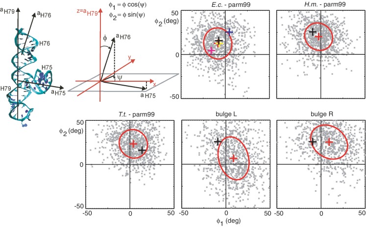
The L1 stalk is a key mobile element of the large ribosomal subunit which interacts with tRNA during translocation. Here, we investigate the structure and mechanical properties of the rRNA H76/H75/H79 three-way junction at the base of the L1 stalk from four different prokaryotic organisms. We propose a coarse-grained elastic model and parameterize it using large-scale atomistic molecular dynamics simulations. Global properties of the junction are well described by a model in which the H76 helix is represented by a straight, isotropically flexible elastic rod, while the junction core is represented by an isotropically flexible spherical hinge. Both the core and the helix contribute substantially to the overall H76 bending fluctuations. The presence of wobble pairs in H76 does not induce any increased flexibility or anisotropy to the helix. The half-closed conformation of the L1 stalk seems to be accessible by thermal fluctuations of the junction itself, without any long-range allosteric effects. Bending fluctuations of H76 with a bulge introduced in it suggest a rationale for the precise position of the bulge in eukaryotes. Our elastic model can be generalized to other RNA junctions found in biological systems or in nanotechnology.


Figures

Figure 1.
Figure 1.
(A and B) Superposition of 23S rRNA in the large ribosomal subunit of E.c. (pdb code 2AW4, red) and T.t. (1VSP, green). The H75/H76/H79 junction at the basis of the L1 stalk is highlighted. Notice the different positions of H76 in the two structures: the E.c. H76 is extended away from the ribosome body (open conformation), the T.t. H76 is tilted towards it (half-closed conformation). The central protuberance (CP) and the L7/L12 stalk are indicated, all the ribosomal proteins and the H77/H78 rRNA at the top of the L1 stalk are omitted for clarity. (A) View from the small subunit, (B) structure rotated through 90° towards the reader. (C and D) Magnified view of the T.t. L1 stalk (pdb code 1VSP) including the H75/H76/H79 junction (atomic and ribbon representation), rRNA helices H77 and H78 (magenta) and the L1 protein (cyan). The L1 protein is not complete in this structure. The orientation in (C and D) is the same as in (A and B), respectively.
Figure 2.
Figure 2.
2D structures of the studied three-way junctions. H75 is in grey, H76 is in green and H79 is in pink. The central UNA/GAN motif is in light yellow and the residues forming a hook-turn are in red. The original X-ray residue numbering is shown. The part of H76 in H.m. which is missing in the X-ray structure was prepared in silico (magenta). Red arrows indicate the two positions where the bulge base (adenine) was introduced.
Figure 3.
Figure 3.
(Diagrams) Schematic representation of the coarse-grained description. The global reference system is defined by the helical axes of H79 and H75, a segment is then chosen within H76. The segment helical axis (aH76) serves to measure the bending magnitude ϕ and the bending direction ψ, from which the global coordinates ϕ12 are computed as shown. (Panels) Scatter plots of ϕ12 for some of the studied systems. Grey points: instantaneous conformations in 100 ps intervals. The coordinate fluctuations are interpreted using a harmonic, anisotropic elastic model. Red crosses: equilibrium conformations, ellipses: energy levels of kBT/2, black crosses: starting X-ray conformations. In the E.c. panel, additional X-ray structures are shown: pdb codes 2I2V (yellow), 3I1N (magenta), 3I1P (blue). The fluctuations are almost isotropic for systems without a bulge. The initial half-closed conformation (T.t., black cross) lies within the kBT/2 contour and is thus thermally accessible. The bulgeL structure observed in eukaryotes shows biologically relevant fluctuations towards the main ribosome body and towards the 30S subunit (compare with the T.t. half-closed starting conformation). Inserting the bulge at the same location but in the opposite strand (bulgeR) results in biologically less relevant movement where H76 stays away from the 30S subunit. Analogous data for the remaining systems are in Supplementary Figure S1.
Figure 4.
Figure 4.
(Left) Schematic illustration of the flexible core, flexible helix model. The coordinates ϕ12 (Figure 3) are decomposed into the contribution from the flexible junction core (ϕJ1J2) and from the flexible helix (ϕH1H2): ϕ1 = ϕJ1 + ϕH1, ϕ2 = ϕJ2 + ϕH2. (Panels) Fitting of the isotropic flexible core, flexible helix model. The contour length is the distance along H76 from its base pair closest to the core to the middle of the segment chosen in H76. The bending angle variation formula image is defined by Equation (6). The model predicts a linear relation between the contour length and the bending angle variation [Equation (5)]. The simulated data (blue crosses) satisfy the linear relation very well. The model stiffness parameters are inferred from the fitted linear functions (red lines). The intersection of the line with the y-axis determines the stiffness of the junction core aJ, the line slope determines the stiffness of the helix expressed by persistence length Lp [Equation (5) and (7)]. The stiffness parameters inferred from the data are shown. The complete list of the inferred parameters is in Table 1, analogous plots for the remaining systems are in Supplementary Figure S3.

References

    1. Rodnina MV, Wintermeyer W, Green R. Ribosomes. Structure, Function, and Dynamics. Springer, Wien, New York. 2011
    1. Korostelev A, Ermolenko DN, Noller HF. Structural dynamics of the ribosome. Curr. Opin. Chem. Biol. 2008;12:674–683. - PMC - PubMed
    1. Schmeing TM, Ramakrishnan V. What recent ribosome structures have revealed about the mechanism of translation. Nature. 2009;461:1234–1242. - PubMed
    1. Blanchard SC. Single-molecule observations of ribosome function. Curr. Opin. Struct. Biol. 2009;19:103–109. - PMC - PubMed
    1. Agirrezabala X, Frank J. Elongation in translation as a dynamic interaction among the ribosome, tRNA, and elongation factors EF-G and EF-Tu. Q. Rev. Biophys. 2009;42:159–200. - PMC - PubMed

Publication types

MeSH terms