. 2012 Apr 24;109(17):6674-9.
doi: 10.1073/pnas.1117412109. Epub 2012 Mar 26.

Molecular MRI enables early and sensitive detection of brain metastases

Affiliations

Molecular MRI enables early and sensitive detection of brain metastases

Sébastien Serres et al. Proc Natl Acad Sci U S A. .

Abstract

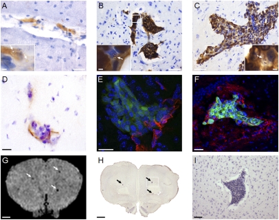
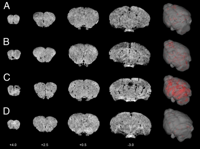
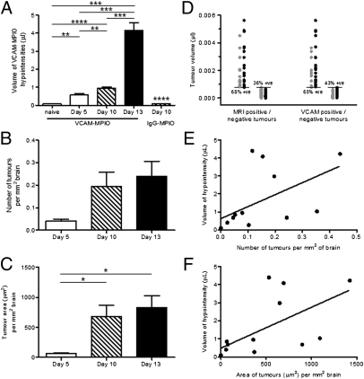
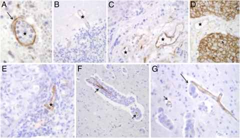
Metastasis to the brain is a leading cause of cancer mortality. The current diagnostic method of gadolinium-enhanced MRI is sensitive only to larger tumors, when therapeutic options are limited. Earlier detection of brain metastases is critical for improved treatment. We have developed a targeted MRI contrast agent based on microparticles of iron oxide that enables imaging of endothelial vascular cell adhesion molecule-1 (VCAM-1). Our objectives here were to determine whether VCAM-1 is up-regulated on vessels associated with brain metastases, and if so, whether VCAM-1-targeted MRI enables early detection of these tumors. Early up-regulation of cerebrovascular VCAM-1 expression was evident on tumor-associated vessels in two separate murine models of brain metastasis. Metastases were detectable in vivo using VCAM-1-targeted MRI 5 d after induction (<1,000 cells). At clinical imaging resolutions, this finding is likely to translate to detection at tumor volumes two to three orders of magnitude smaller (0.3-3 × 10(5) cells) than those volumes detectable clinically (10(7)-10(8) cells). VCAM-1 expression detected by MRI increased significantly (P < 0.0001) with tumor progression, and tumors showed no gadolinium enhancement. Importantly, expression of VCAM-1 was shown in human brain tissue containing both established metastases and micrometastases. Translation of this approach to the clinic could increase therapeutic options and change clinical management in a substantial number of cancer patients.


Conflict of interest statement

The authors declare no conflict of interest.

Figures

Fig. 1.
Fig. 1.
Immunohistochemical detection of brain metastases in 4T1 model. (A–C) Photomicrographs of tumor colonies at days 5 (A), 10 (B), and 13 (C) after 4T1 injection. (Scale bars: 100 μm.) Insets show magnified regions of the tumors with endothelial binding of VCAM–MPIO (arrows). (Scale bars: 20 μm.) (D) Immunohistochemical colocalization of VCAM-1 (brown) with a brain metastasis at day 10. (E and F) Colocalization of VCAM-1 (red) with GFP-positive tumors (green) detected by confocal microscopy at days 10 and 13, respectively. Cell nuclei are stained blue. (Scale bars: D–F, 10 μm.) (G–I) Colocalization of focal hypointensities on a T2*-weighted image (G; arrows) with histological detection of metastases (H; arrows). (I) Zoomed view of the box indicated in H. (Scale bars: G and H, 1 mm; I, 100 μm.)
Fig. 2.
Fig. 2.
MRI detection of VCAM–MPIO binding in 4T1 model. Selected T2*-weighted images from a 3D dataset (coordinates relative to Bregma) at days 5 (A), 10 (B), and 13 (C) after intracardiac injection of 4T1 cells. Intense focal hypointense areas (black) correspond to MPIO binding. No specific MPIO binding was detected in naïve BALB/c mice injected with VCAM–MPIO (D). 3D reconstructions (column 5) show the spatial distribution of VCAM–MPIO binding (red) throughout the brain.
Fig. 3.
Fig. 3.
Quantitation of VCAM–MPIO binding and tumor number/area in 4T1 model. (A) The volume of hypointensities on T2*-weighted images in VCAM–MPIO-injected animals increased significantly over time (n = 5–6 per group). Significance shown for the day 10 IgG–MPIO group is compared with the day 10 VCAM–MPIO group. (B and C) Graphs to show change in number (B) and area (C) of tumor colonies over time after intracardiac 4T1 cell injection (n = 3 on day 5; n = 4 on days 10 and 13). (D) Graph indicating volumes of metastases at day 10 that were MRI- or VCAM-1–positive (black circles) and MRI- or VCAM-1–negative (gray circles). Percentages of positive values (MRI or VCAM-1) are given for cohorts of tumors above and below the mean tumor volume (horizontal lines). Correlation analysis between the volume of hypointensities on T2*-weighted images and the number (E) or area (F) of tumor colonies revealed significant positive correlations (P < 0.05; r2 = 0.3 and 0.4, respectively). In all cases, *P < 0.05, **P < 0.01, ***P < 0.001, and ****P < 0.0001.
Fig. 4.
Fig. 4.
VCAM–MPIO binding and tumor detection in the MDA231BR model. (A) Selected T2*-weighted images from a 3D dataset (coordinates relative to Bregma) at day 21 after intracardiac injection of MDA231BR cells. Intense focal hypointense areas (black) correspond to VCAM–MPIO binding (Scale bar: 1 mm.) (B and C) Colocalization of the MRI hypointense signals (arrows) with VCAM-1 expression (brown) and metastases (arrows). C1–C4 show higher magnification photomicrographs of the four tumor colonies highlighted in C. (Scale bars: B, 1 mm; C, 200 μm; C1–C4, 50 μm.) (D) 3D reconstruction shows the spatial distribution of VCAM–MPIO binding (red).
Fig. 5.
Fig. 5.
VCAM-1 expression in human brain metastasis. (A) Positive control section showing VCAM-1 expression (brown) on a brain vessel (★) adjacent to acute inflammation. (B) Normal brain tissue showing minimal VCAM-1 reaction in cortical vessel (★). (C) Strong VCAM-1 staining in stromal endothelial cells of a metastatic carcinoma (★); solid carcinoma is in the upper left corner. (D) In one case, VCAM-1 staining was evident on the membranes of the carcinoma cells themselves as well as on stromal vessels (★). (E) Substantial endothelial VCAM-1 expression in a stromal vessel of a metastasis that evoked chronic inflammation (★). (F and G) Selective expression of VCAM-1 by endothelial cells in close proximity to perivascular brain micrometastases (arrows). Note intimate association of three tumor cells (G; long arrow) with a VCAM-1–positive small vessel. (Magnification: A–F, 400×; G; 200×.)

References

    1. Nussbaum ES, Djalilian HR, Cho KH, Hall WA. Brain metastases. Histology, multiplicity, surgery, and survival. Cancer. 1996;78:1781–1788. - PubMed
    1. Weil RJ, Palmieri DC, Bronder JL, Stark AM, Steeg PS. Breast cancer metastasis to the central nervous system. Am J Pathol. 2005;167:913–920. - PMC - PubMed
    1. Hochstenbag MM, Twijnstra A, Wilmink JT, Wouters EF, ten Velde GP. Asymptomatic brain metastases (BM) in small cell lung cancer (SCLC): MR-imaging is useful at initial diagnosis. J Neurooncol. 2000;48:243–248. - PubMed
    1. Suzuki K, et al. Magnetic resonance imaging and computed tomography in the diagnoses of brain metastases of lung cancer. Lung Cancer. 2004;46:357–360. - PubMed
    1. Krüger S, et al. Brain metastasis in lung cancer. Comparison of cerebral MRI and 18F-FDG-PET/CT for diagnosis in the initial staging. Nucl Med (Stuttg) 2011;50:101–106. - PubMed

Publication types

Substances