. 2012 Apr 10;109(15):5803-8.
doi: 10.1073/pnas.1202922109. Epub 2012 Mar 26.

Mutant induced pluripotent stem cell lines recapitulate aspects of TDP-43 proteinopathies and reveal cell-specific vulnerability

Affiliations

Mutant induced pluripotent stem cell lines recapitulate aspects of TDP-43 proteinopathies and reveal cell-specific vulnerability

Bilada Bilican et al. Proc Natl Acad Sci U S A. .

Abstract

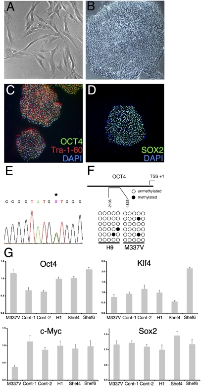
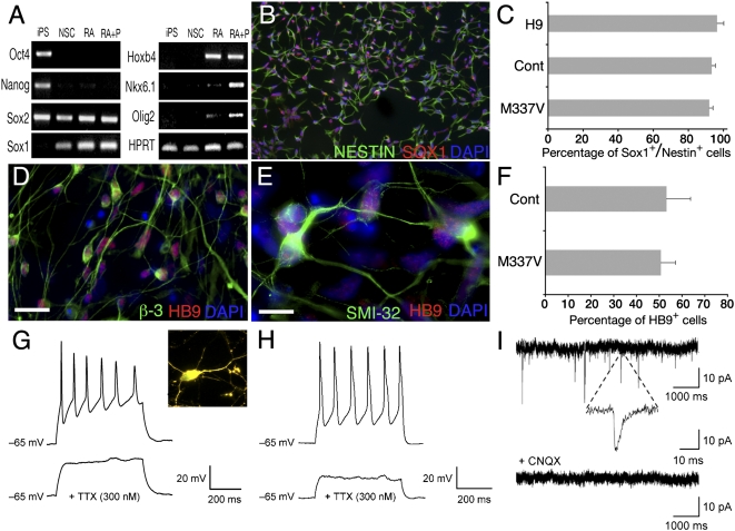
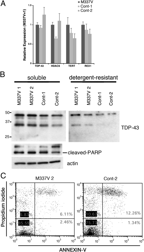
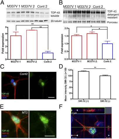
Transactive response DNA-binding (TDP-43) protein is the dominant disease protein in amyotrophic lateral sclerosis (ALS) and a subgroup of frontotemporal lobar degeneration (FTLD-TDP). Identification of mutations in the gene encoding TDP-43 (TARDBP) in familial ALS confirms a mechanistic link between misaccumulation of TDP-43 and neurodegeneration and provides an opportunity to study TDP-43 proteinopathies in human neurons generated from patient fibroblasts by using induced pluripotent stem cells (iPSCs). Here, we report the generation of iPSCs that carry the TDP-43 M337V mutation and their differentiation into neurons and functional motor neurons. Mutant neurons had elevated levels of soluble and detergent-resistant TDP-43 protein, decreased survival in longitudinal studies, and increased vulnerability to antagonism of the PI3K pathway. We conclude that expression of physiological levels of TDP-43 in human neurons is sufficient to reveal a mutation-specific cell-autonomous phenotype and strongly supports this approach for the study of disease mechanisms and for drug screening.


Conflict of interest statement

The authors declare no conflict of interest.

Figures

Fig. 1.
Fig. 1.
Establishment of iPSCs from patient fibroblasts. (A and B) Primary dermal fibroblasts from an ALS patient (A) formed tightly packed hESC-like colonies (B) after lentiviral reprogramming with OCT4, SOX2, KLF4, and c-MYC. (C and D) Immunohistochemical characterization of feeder-free patient iPSCs for the pluripotency markers OCT4, Tra-1-60, and SOX2. (E) Direct sequencing confirmed the M337V mutation in TDP-43 in patient-derived iPSCs. (F) Bisulfite sequencing analysis of the OCT4 promoter in M337V iPSCs and hESC line H9s. (G) qRT-PCR comparison of total expression levels of OCT4, KLF4, c-MYC, and SOX2 in M337V and WT iPSCs and hESC lines H1, Shef4, and Shef6. Values are mean ± SEM. All results are from M337V iPSC clone 2 (M337V-2) except G and are representative of iPSC clone M337V-1.
Fig. 2.
Fig. 2.
TDP-43 M337V and control iPSCs can be differentiated into functional MNs. iPSCs or hESCs were directed toward the neuroectodermal lineage by dual-SMAD signaling inhibition in feeder-free, adherent culture conditions. (A) M337V iPSCs down-regulate OCT4 and NANOG expression in response to neuralization and up-regulate the lineage-specific genes SOX1, HOXB4, NKX6.1, and OLIG2 in a temporal order after sequential application of MN patterning factors retinoic acid (RA) and purmorphamine (P). (B) Neural differentiation was assessed by quantitative immunohistochemical costaining for Nestin and Sox1 after replating. (C) M337V and control iPSCs and H9 hESCs showed similar efficiency in neural differentiation. (D and E) At 4–6 wk of maturation, iPSC-derived MN progenitors plated on laminin/fibronectin express HB9 and β3-tubulin (D) and HB9 and SMI-32 (E). (F) M337V and control iPSC-derived neural stem cells (NSCs) generated MNs with equal efficiency, as assessed by quantitative immunohistochemistry for HB9. (G and H) Current–clamp traces showed tetrodotoxin (TTX)-sensitive action potentials in response to injection of depolarizing current in M337V (G) and WT (H) MNs. (G Inset) ChAT and LcY double-positive neuron from which recording is performed. (I) Sample traces of spontaneous miniature EPSCs recorded from an M337V culture. All miniature EPSC events were blocked with CNQX. All data are from M337V-2 and Cont-2 iPSCs. Values are mean ± SEM (n = 3). (Scale bars: 10 μm.)
Fig. 3.
Fig. 3.
M337V iPSCs display biochemical features of TDP-43 misaccumulation. (A) M337V and control iPSCs express similar levels of TDP-43, REX1, TERT, and HDAC6, as determined by qRT-PCR. (B) Immunoblots of the detergent-soluble and -insoluble fractions from feeder-free iPSC lysates. TDP-43 fragments of ∼35 kDa are enriched in the detergent-resistant fraction of the M337V iPSC lysates. Soluble or insoluble TDP-43 levels do not correlate with cleaved PARP. β-Actin served as a loading control. (C) Representative annexin V/propidium iodide staining by flow cytometry. The percentages of cells negative for both annexin V and propidium iodide were similar in M337V-2 (85.6%) and Cont-2 (75.3%) iPSCs, suggesting comparable levels of cell death under basal conditions.
Fig. 4.
Fig. 4.
M337V neurons have higher levels of soluble and detergent-resistant TDP-43. (A and B) Western blot analysis of M337V-1, M337V-2, and Cont-2 MN-containing cultures. Three independent extractions are shown for each cell line. β3-Tubulin was the loading control for soluble fractions (A), and Ponceau S reversible membrane staining (51) was the loading control for detergent-resistant fractions (B). Semiquantitative analyses of band signal intensity was accomplished by using ImageJ. (A) Levels of soluble TDP-43 normalized to β3-tubulin, Cont-2 set to 1: M337V-1 = 3.67 ± 0.58; M337V-2 = 4.54 ± 0.01; Cont-2 = 1.0 ± 0.24. M337V-1 vs. M337V-2, not significant; M337V-1 vs. Cont-2, P = 0.013; M337V-2 vs. Cont-2, P < 0.001, n = 3 independent extractions. (B) Levels of detergent-resistant TDP-43 normalized to Ponceau S, Cont-2 set to 1: M337V-1 = 1.52 ± 0.08; M337V-2= 1.67 ± 0.10; Cont-2 = 1.0 ± 0.16. M337V-1 vs. M337V-2, not significant; M337V-1 vs. Cont-2, P = 0.04; M337V-2 vs. Cont-2, P < 0.03, n = 3 independent extractions. Values are mean ± SEM. Data were analyzed by one-way ANOVA and post hoc Tukey test. (C and D) Immunofluorescence analysis of nuclear TDP-43 in SMI-32+ and SM-I32 cells. Cont-2 SMI-32+ (118.89% ± 2.6%) vs. SMI-32 (100% ± 2.9%), P < 0.001, n = 40 cells for each group. (E) M337V iPSC-derived SMI-32+ MNs displayed predominantly nuclear TDP-43 localization with granular staining present in soma and neurites. (F) Orthogonal views through one TDP-43 M337V SMI-32+ cell demonstrating localization of TDP-43 (green), SMI-32 (red), and DAPI (blue). (Scale bars: 10 μm.)
Fig. 5.
Fig. 5.
M337V TDP-43 neurons display selective vulnerability. (A) Representative image of an HB9::GFP+ neuron used in real-time survival analysis. (B) Real-time survival analysis of M337V-1, M337V-2, Cont-1, and Cont-2 MN-containing cultures showing cumulative risk of death of HB9::GFP-transfected neurons. Pooled data from three independent differentiation experiments are shown (P < 0.001 between mutant and control groups, log-rank test). (C) Cytotoxicity in MN cultures derived from M337V and control iPSCs after treatment with LY294002 (40 μM) for 48 h. A total of 20,000 MN precursors per well were plated in a 96-well format and allowed to differentiate for 4 wk before treatments were started. Fluorescent LDH release measured with a FLUOstar OPTIMA plate reader served as a measure of cytotoxicity. Released LDH was normalized to total LDH for each well, and mock-treated samples were set to 100%. Percentage cytotoxicity for each treatment was determined as a factor of mock treatment for each cell line. Values are mean ± SEM. Data were analyzed by one-way ANOVA and post hoc Tukey test. *P < 0.05; M337V-1 vs. M337V-2, not significant; n = 3 independent experiments.

References

    1. Neumann M, et al. Ubiquitinated TDP-43 in frontotemporal lobar degeneration and amyotrophic lateral sclerosis. Science. 2006;314(5796):130–133. - PubMed
    1. Higashi S, et al. Concurrence of TDP-43, tau and α-synuclein pathology in brains of Alzheimer's disease and dementia with Lewy bodies. Brain Res. 2007;1184:284–294. - PubMed
    1. Arai T, et al. TDP-43 is a component of ubiquitin-positive tau-negative inclusions in frontotemporal lobar degeneration and amyotrophic lateral sclerosis. Biochem Biophys Res Commun. 2006;351:602–611. - PubMed
    1. Uryu K, et al. Concomitant TAR-DNA-binding protein 43 pathology is present in Alzheimer disease and corticobasal degeneration but not in other tauopathies. J Neuropathol Exp Neurol. 2008;67:555–564. - PMC - PubMed
    1. Yokoseki A, et al. TDP-43 mutation in familial amyotrophic lateral sclerosis. Ann Neurol. 2008;63:538–542. - PubMed

Publication types

MeSH terms