. 2012 Apr 10;109(15):5874-9.
doi: 10.1073/pnas.1200447109. Epub 2012 Mar 26.

Dichotomous effects of VEGF-A on adipose tissue dysfunction

Affiliations

Dichotomous effects of VEGF-A on adipose tissue dysfunction

Kai Sun et al. Proc Natl Acad Sci U S A. .

Abstract

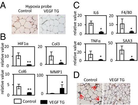
Obese fat pads are frequently undervascularized and hypoxic, leading to increased fibrosis, inflammation, and ultimately insulin resistance. We hypothesized that VEGF-A-induced stimulation of angiogenesis enables sustained and sufficient oxygen and nutrient exchange during fat mass expansion, thereby improving adipose tissue function. Using a doxycycline (Dox)-inducible adipocyte-specific VEGF-A overexpression model, we demonstrate that the local up-regulation of VEGF-A in adipocytes improves vascularization and causes a "browning" of white adipose tissue (AT), with massive up-regulation of UCP1 and PGC1α. This is associated with an increase in energy expenditure and resistance to high fat diet-mediated metabolic insults. Similarly, inhibition of VEGF-A-induced activation of VEGFR2 during the early phase of high fat diet-induced weight gain, causes aggravated systemic insulin resistance. However, the same VEGF-A-VEGFR2 blockade in ob/ob mice leads to a reduced body-weight gain, an improvement in insulin sensitivity, a decrease in inflammatory factors, and increased incidence of adipocyte death. The consequences of modulation of angiogenic activity are therefore context dependent. Proangiogenic activity during adipose tissue expansion is beneficial, associated with potent protective effects on metabolism, whereas antiangiogenic action in the context of preexisting adipose tissue dysfunction leads to improvements in metabolism, an effect likely mediated by the ablation of dysfunctional proinflammatory adipocytes.


Conflict of interest statement

The authors declare no conflict of interest.

Figures

Fig. 1.
Fig. 1.
VEGF-A stimulates angiogenesis in WAT. (A) Functional blood vessels in SWAT and BAT visualized by tail-injected rhodamine tagged lectin-1 in VEGF-A Tg and control mice. Blood vessels are shown in red and the nuclei are in blue, visualized by DAPI staining. The images were captured with confocal microscope. (B) q-PCR analysis of CD31 in SWAT of VEGF-A Tg and control mice (n = 4 in controls; n = 5 in VEGF-A Tg). The difference was analyzed by Student's t test. *P < 0.05. (C) Immunohistochemical analysis with α-CD31 in SWAT of VEGF Tg or their littermate controls. (Scale bar, 50 μm.) (D) Immunohistochemical analysis with α-VEGF receptor 2 and α-phosphorylated VEGF receptor 2 in SWAT in both VEGF Tg and controls. (Scale bar, 50 μm.)
Fig. 2.
Fig. 2.
VEGF-A Tg mice exhibit an improved metabolic profile. (A) Circulating glucose levels measured during an OGTT (n = 5 per group) 5 wk after HFD feeding. The difference at each time point was analyzed by Student's t test. *P < 0.05; **P < 0.001. (B) Indirect calorimetry was performed in a CLAMS system by housing mice after HFD plus Dox feeding for 6 wk. VO2 and RER (VCO2/VO2) were calculated from the average results during a 24-h light and dark cycle. Absolute contributions of carbohydrate and lipid metabolism to total energy expenditure and accumulated food intake were measured (ACC). *P < 0.05; **P < 0.001.
Fig. 3.
Fig. 3.
VEGF-A Tg mice have smaller white adipocytes with BAT-like properties. (A) H&E staining of SWAT of VEGF-A Tg mice after HFD plus Dox feeding for 8 wk. (Scale bars, 50 μm.) (B) q-PCR analysis of PGC-1α and UCP-1 in EWAT of VEGF-A Tg and their littermate controls. The readings are normalized to hypoxanthine phosphoribosyltransferase (HPRT). *P < 0.05; **P < 0.001. (C) Western blot analysis for both PGC-1α and UCP-1 in WAT of VEGF Tg and their littermate controls. Results were normalized with β-actin.
Fig. 4.
Fig. 4.
VEGF-A overexpression ameliorates hypoxia, fibrosis, and inflammatory responses in WAT induced by HFD. (A) Immunohistochemical staining by hypoprobe-1 in WAT of VEGF Tg mice and their littermate controls after HFD plus Dox treatment for 8 wk. The darker staining on the Left indicates more hypoxic environment in controls. (B) q-PCR analysis of HIF-1α and its target collagen genes in EWAT. The readings are normalized to HPRT. *P < 0.05; **P < 0.001. (C) q-PCR analysis of inflammatory cytokines IL6, TNFα, SAA3, and macrophage marker F4/80 in EWAT. The readings are normalized to HPRT. *P < 0.05; **P < 0.001. (D) Immunohistochemical staining of F4/80 in SWAT of VEGF-A Tg mice. Red arrows indicate the crowns formed by accumulation of macrophages surrounding the dysfunctional adipocytes.
Fig. 5.
Fig. 5.
Blockade of VEGF-A binding to VEGFR2 by Mcr84 accelerates metabolic dysfunction induced by HFD. (A) Functional blood vessels in AT labeled by rhodamine-tagged lectin. The mice were fed with HFD for 6 wk before the experiment. Mcr84 or control IgG were injected i.p at the initial stage and continued twice per week for the whole HFD feeding process. Blood vessels are shown in red and the nucleus in blue, by DAPI staining. (B) Insulin levels for an OGTT in Mcr84- or control IgG-treated mice (n = 5 per group) after antibody treated for 6 wk. *P < 0.05. (C) Triglyceride levels for a lipid clearance test in Mcr84- or control IgG-treated mice (n = 5 per group). *P < 0.05. (D) Serum nonesterified fatty acid (NEFA) levels in Mcr84- or control IgG-treated mice (n = 5 per group) after antibody treated for 7 wk. *P < 0.05.

References

    1. Rosen ED, MacDougald OA. Adipocyte differentiation from the inside out. Nat Rev Mol Cell Biol. 2006;7:885–896. - PubMed
    1. Sethi JK, Vidal-Puig AJ. Thematic review series: Adipocyte biology. Adipose tissue function and plasticity orchestrate nutritional adaptation. J Lipid Res. 2007;48:1253–1262. - PMC - PubMed
    1. Sun K, Kusminski CM, Scherer PE. Adipose tissue remodeling and obesity. J Clin Invest. 2011;121:2094–2101. - PMC - PubMed
    1. Cao Y. Adipose tissue angiogenesis as a therapeutic target for obesity and metabolic diseases. Nat Rev Drug Discov. 2010;9:107–115. - PubMed
    1. Zhang QX, et al. Vascular endothelial growth factor is the major angiogenic factor in omentum: Mechanism of the omentum-mediated angiogenesis. J Surg Res. 1997;67:147–154. - PubMed

Publication types

MeSH terms

Substances

LinkOut - more resources