. 2012 Apr 10;109(15):5645-50.
doi: 10.1073/pnas.1120269109. Epub 2012 Mar 26.

Calcium and pH-dependent packing and release of the gel-forming MUC2 mucin

Affiliations

Calcium and pH-dependent packing and release of the gel-forming MUC2 mucin

Daniel Ambort et al. Proc Natl Acad Sci U S A. .

Abstract

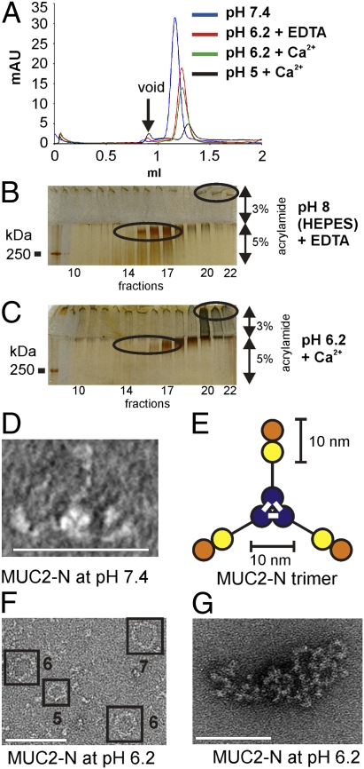
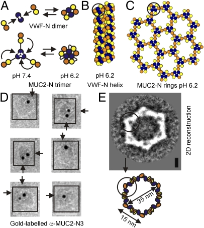
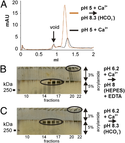
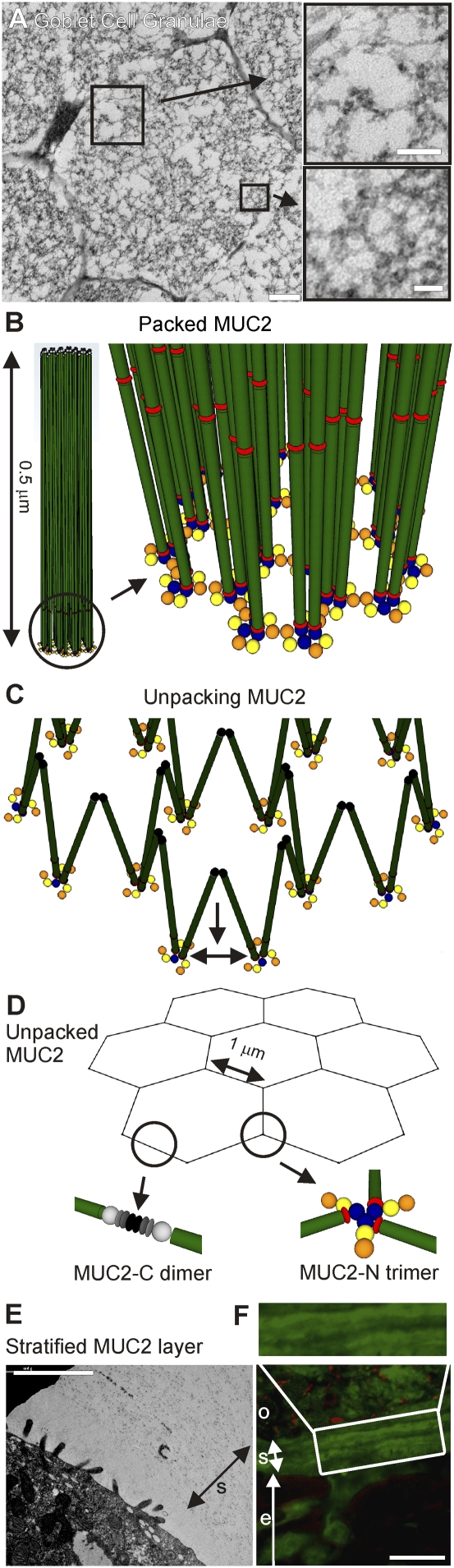
MUC2, the major colonic mucin, forms large polymers by N-terminal trimerization and C-terminal dimerization. Although the assembly process for MUC2 is established, it is not known how MUC2 is packed in the regulated secretory granulae of the goblet cell. When the N-terminal VWD1-D2-D'D3 domains (MUC2-N) were expressed in a goblet-like cell line, the protein was stored together with full-length MUC2. By mimicking the pH and calcium conditions of the secretory pathway we analyzed purified MUC2-N by gel filtration, density gradient centrifugation, and transmission electron microscopy. At pH 7.4 the MUC2-N trimer eluted as a single peak by gel filtration. At pH 6.2 with Ca(2+) it formed large aggregates that did not enter the gel filtration column but were made visible after density gradient centrifugation. Electron microscopy studies revealed that the aggregates were composed of rings also observed in secretory granulae of colon tissue sections. The MUC2-N aggregates were dissolved by removing Ca(2+) and raising pH. After release from goblet cells, the unfolded full-length MUC2 formed stratified layers. These findings suggest a model for mucin packing in the granulae and the mechanism for mucin release, unfolding, and expansion.


Conflict of interest statement

The authors declare no conflict of interest.

Figures

Fig. 1.
Fig. 1.
Domain structure of MUC2 and N-terminal protein and storage of MUC2 N terminus in goblet cell vesicles. (A) MUC2 monomer consists of multiple domains: VWD [D1 (orange), D2 (yellow), D′D3 (blue) and D4 (light gray)], CysD (red), PTS (green), VWB (gray), VWC (dark gray), and cystine-knot CK (black). The MUC2-N plasmid contains the N-terminal VWD (D1-D2-D′D3) domains, CysD1, Myc-tag (black), and GFP (light green). The α-MUC2-N3 antibody recognizes the VWD′D3 domain. (B) Differentiated LS174TdnTCF4 cells expressing MUC2-N (green) were immunostained with anti-MUC2C2m1 MAb (red) detecting endogenous MUC2 showing colocalization. (Scale bars, 5 μm.) (C) LS174TdnTCF4 cells expressing the MUC2-N protein (green) were induced to differentiate into goblet-like cells. Increasing accumulation of MUC2-N in larger vesicles was observed in the more differentiated cells. Nucleus, DAPI (blue). (D and E) Induced LS174TdnTCF4 with MUC2-N (green) were immunostained for ER with anti-calnexin (red) (D) or for Golgi with anti-GalNAcT2 (red) (E). No colocalization was apparent. (Scale bars, 5 μm.) (F) Lysate from a radioactive pulse-chase experiment (30 min, 3 h, 20 h) of LS174TdnTCF4 (noninduced) expressing the MUC2-N protein that was immunoprecipitated using anti-myc MAb and analyzed by nonreduced SDS/PAGE. MUC2-N is first detected as a monomer, and during processing when passing the secretory pathway it forms trimers that were stored within the cells for more than 20 h.
Fig. 2.
Fig. 2.
MUC2-N forms pH- and calcium-dependent rings. (A) Gel filtration of MUC2-N at pH 7.4, pH 6.2 with EDTA or Ca2+, and pH 5 with Ca2+. The MUC2-N trimer formed larger aggregates at lower pH that did not enter the gel beads. (B and C) Density gradient centrifugation of MUC2-N protein incubated at pH 8 with EDTA (B) or pH 6.2 with Ca2+ (C). As analyzed by reducing SDS/PAGE and silver staining, the MUC2-N trimer was found in fractions 14–18 in high and low pH conditions, whereas larger aggregates were detected in fractions 20–22 only at pH 6.2 with Ca2+. (D) EM (negative stain) showing that MUC2-N trimer adopts an outstretched appearance at pH 7.4, with three globular structures extending out of a trefoil core fragment, suggesting that the VWD1-D2 domains are connected via a flexible linker to the disulfide-bonded (indicated by white bars) VWD3 trimeric core (E) (same color code as in Fig. 1). (F) EM showing that MUC2-N forms five-, six-, and seven-sided ring structures at pH 6.2 with Ca2+ composed of multiple MUC2-N trimers. (G) Glutaraldehyde cross-linking at pH 6.2 with Ca2+ before density gradient centrifugation (fraction 22 from C) allowed for isolating confluent 2D sheets of concatenated MUC2-N rings as seen by EM (negative stain). (Scale bars, 50 nm in D, 100 nm in F and G.)
Fig. 3.
Fig. 3.
Domain organization of MUC2-N six-sided rings. (A) The MUC2-N trimer is made up of three disulfide-linked (white bars) VWD3 (blue) domains in the core structure. At pH 7.4 the VWD1-D2 (orange and yellow) domains extend out and at pH 6.2 with Ca2+ flipped back by noncovalent interactions to the VWD3 core (Lower). In VWF the N-terminal VWD′D3 domains form a disulfide-linked (white bars) dimer. At pH 6.2 two VWD1-D2 domains interact with the VWD3 dimer, thus forming a tetrameric complex (Upper). (B) At pH 6.2 with Ca2+ in the VWF-N two disulfide-linked VWD3 domains bind on each side one VWD1-D2 peptide thereby forming the repeating unit of a right-handed helix. The VWF-N helix is composed of four repeating units per turn. (C) Analogously, the MUC2-N repeating unit is composed of three disulfide-linked VWD3 domains that bind on each side one VWD1-D2 peptide. Six repeating units make up one ring. (D) EM (negatively stained) showing the gold-labeled α-MUC2-N3 antibodies for the VWD3 domains bound to the corners of six different MUC2-N rings formed at pH 6.2 with Ca2+ (each arrow shows one labeled corner). (E) 2D reconstruction of MUC2-N six-sided rings by EM shows a six-folded symmetry, thereby suggesting six repeating units per ring. Each repeating unit is made up of three VWD3 domains and three VWD1-D2 peptides. (Scale bars, 40 nm in D, 7.5 nm in E.)
Fig. 4.
Fig. 4.
Dissolution of MUC2-N aggregates by Hepes-EDTA or NaHCO3-treatment. (A) MUC2-N aggregates formed at pH 5 with 50 mM CaCl2 were analyzed by gel filtration with or without addition of 100 mM sodium bicarbonate (pH 8.3). Addition of sodium bicarbonate partially dissolved the MUC2-N aggregates. (B and C) MUC2-N aggregates formed at pH 6.2 with Ca2+ were dialyzed against 20 mM Hepes (pH 8), 10 mM EDTA (B) or 50 mM NaHCO3 (pH 8.3) (C) and then analyzed by density gradient centrifugation and reducing SDS/PAGE and silver staining. Higher pH conditions or sodium bicarbonate treatment both partially dissolved the MUC2-N aggregates that were formed at low pH with Ca2+ in fractions 20–22.
Fig. 5.
Fig. 5.
Packing and unfolding of MUC2. (A and B) Packing of MUC2 at pH 6.2 in presence of Ca2+ in secretory granulae of goblet cell. (A) EM of a cross-section of secretory granulae from a human goblet cell showing ring-like structures. (Scale bars, 200 nm at left, 100 nm at top right, 40 nm at bottom right.) (B) MUC2 is organized at one end around the MUC2-N concatenated rings stabilized by noncovalent interactions of N-terminal VWD (orange, yellow, and blue) domains and at the C-terminal end by CK (black) dimers. The extended mucin domains drawn to scale (green) connect neighboring VWD′D3 trimers (blue) of the MUC2-N ring platform (Lower) with the C-terminal ends (Upper). (C and D) Unfolding of MUC2 at high pH and low Ca2+ after release from goblet cell secretory granulae. (C) During unpacking the MUC2-N rings fall apart, and the mucin domains begin to stretch out. (D) The released MUC2 is assumed to adopt an outstretched net-like flat sheet composed of trimeric disulfide-linked MUC2-N (in the corners) and dimeric MUC2-C (in the middle of the sides). The expanded net is >1,000-fold larger than the granulae rings. (E and F) The inner mucus layer of the colon show stratified sheets (5) as predicted from the unfolded MUC2 model. (E) EM of the inner mucus layer organized as stratified sheets (s) on top of an enterocyte with its glycocalyx from a colon tissue section. (Scale bar, 2 μm.) (F) Mouse colon tissue section fixed in Carnoy and stained with the anti-MUC2C3 antiserum (green) and FISH (red) as revealed by fluorescence microscopy of the stratified MUC2 inner mucus layer (s) devoid of bacteria and the outer mucus layer (o) with bacteria. (Scale bar, 25 μm.)

Comment in

  • Mucus supramolecular topology: an elusive riddle.
    Verdugo P. Verdugo P. Proc Natl Acad Sci U S A. 2012 Oct 30;109(44):E2956; author reply E2957. doi: 10.1073/pnas.1211117109. Epub 2012 Sep 17. Proc Natl Acad Sci U S A. 2012. PMID: 22988128 Free PMC article. No abstract available.

Similar articles

Cited by

References

    1. Thornton DJ, Rousseau K, McGuckin MA. Structure and function of the polymeric mucins in airways mucus. Annu Rev Physiol. 2008;70:459–486. - PubMed
    1. Johansson MEV, et al. Composition and functional role of the mucus layers in the intestine. Cell Mol Life Sci. 2011;68:3635–3641. - PMC - PubMed
    1. Lang T, Hansson GC, Samuelsson T. Gel-forming mucins appeared early in metazoan evolution. Proc Natl Acad Sci USA. 2007;104:16209–16214. - PMC - PubMed
    1. Gum JR, Jr, Hicks JW, Toribara NW, Siddiki B, Kim YS. Molecular cloning of human intestinal mucin (MUC2) cDNA. Identification of the amino terminus and overall sequence similarity to prepro-von Willebrand factor. J Biol Chem. 1994;269:2440–2446. - PubMed
    1. Johansson ME, et al. The inner of the two Muc2 mucin-dependent mucus layers in colon is devoid of bacteria. Proc Natl Acad Sci USA. 2008;105:15064–15069. - PMC - PubMed

Publication types

LinkOut - more resources