. 2012 Apr 18;134(15):6625-36.
doi: 10.1021/ja210588m. Epub 2012 Apr 6.

Bifunctional compounds for controlling metal-mediated aggregation of the aβ42 peptide

Affiliations

Bifunctional compounds for controlling metal-mediated aggregation of the aβ42 peptide

Anuj K Sharma et al. J Am Chem Soc. .

Abstract

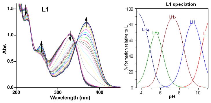
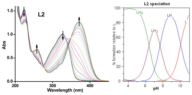
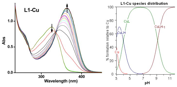
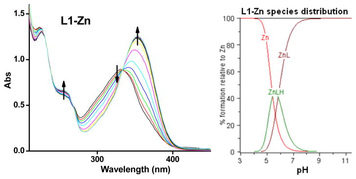
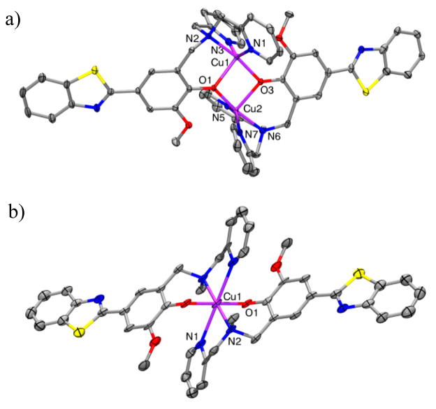
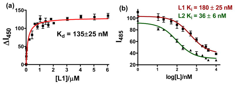
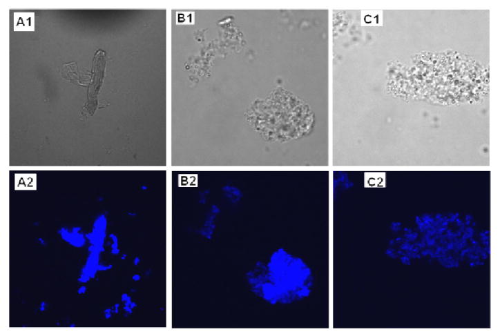
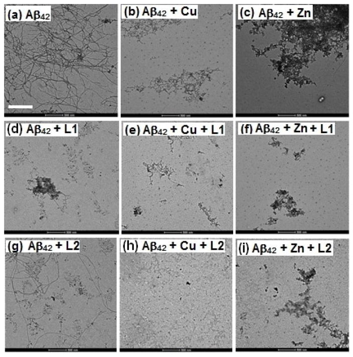
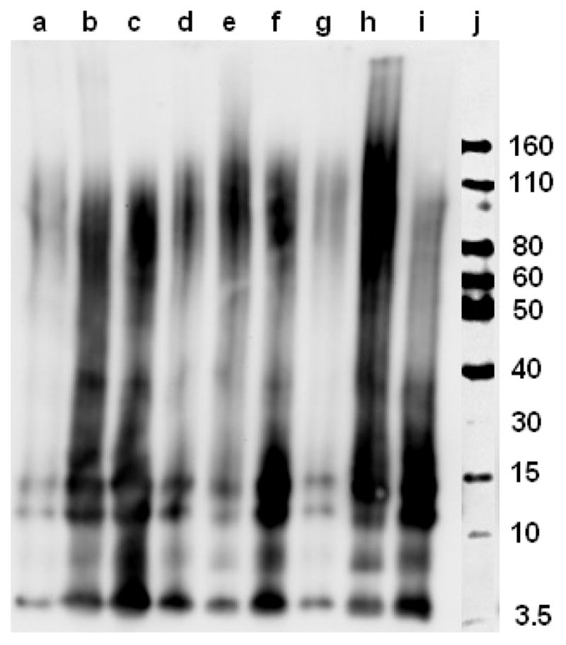
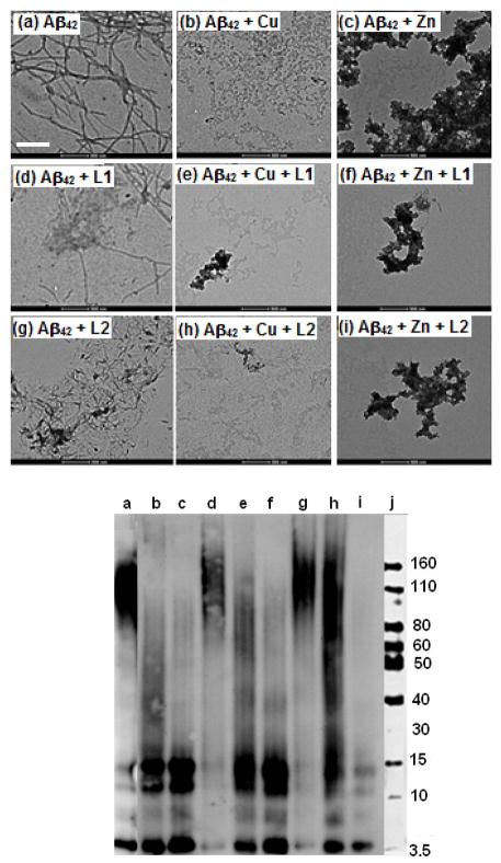
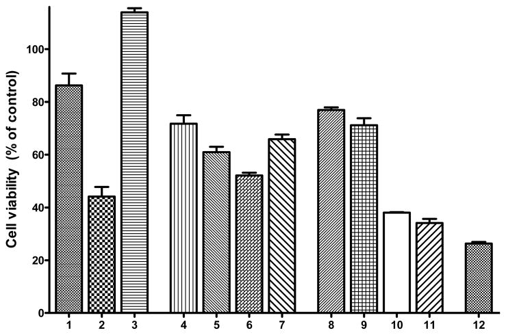
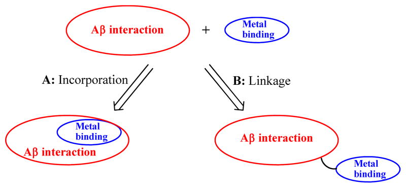
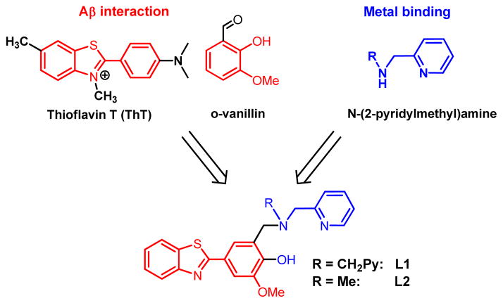
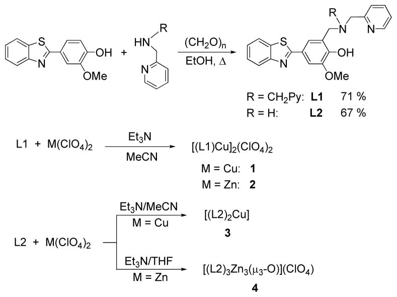
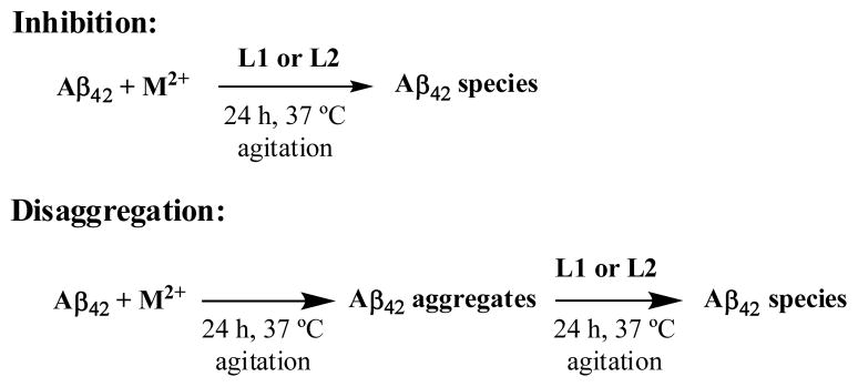
Abnormal interactions of Cu and Zn ions with the amyloid β (Aβ) peptide are proposed to play an important role in the pathogenesis of Alzheimer's disease (AD). Disruption of these metal-peptide interactions using chemical agents holds considerable promise as a therapeutic strategy to combat this incurable disease. Reported herein are two bifunctional compounds (BFCs) L1 and L2 that contain both amyloid-binding and metal-chelating molecular motifs. Both L1 and L2 exhibit high stability constants for Cu(2+) and Zn(2+) and thus are good chelators for these metal ions. In addition, L1 and L2 show strong affinity toward Aβ species. Both compounds are efficient inhibitors of the metal-mediated aggregation of the Aβ(42) peptide and promote disaggregation of amyloid fibrils, as observed by ThT fluorescence, native gel electrophoresis/Western blotting, and transmission electron microscopy (TEM). Interestingly, the formation of soluble Aβ(42) oligomers in the presence of metal ions and BFCs leads to an increased cellular toxicity. These results suggest that for the Aβ(42) peptide-in contrast to the Aβ(40) peptide-the previously employed strategy of inhibiting Aβ aggregation and promoting amyloid fibril dissagregation may not be optimal for the development of potential AD therapeutics, due to formation of neurotoxic soluble Aβ(42) oligomers.


Figures

Figure 1
Figure 1
Variable pH UV spectra of L1 ([L1] = 50 μM, 25 °C, I = 0.1 M NaCl) and species distribution plot.
Figure 2
Figure 2
Variable pH (pH 3–11) UV spectra of L2 ([L2] = 50 μM, 25 °C, I = 0.1 M NaCl) and species distribution plot.
Figure 3
Figure 3
Variable pH (pH 3–11) UV spectra of L1 and Cu2+ system ([L1] = [Cu2+] = 50 μM, 25 °C, I = 0.1 M NaCl) and species distribution plot.
Figure 4
Figure 4
Variable pH (pH 3–11) UV spectra of L1 and Zn2+ ([L1] = [Zn2+] = 50 μM, 25 °C, I = 0.1 M NaCl) and species distribution plot.
Figure 5
Figure 5
ORTEP view of (a) the dication of 1 and (b) 3 with 50% probability ellipsoids. All hydrogen atoms, counteranions, and solvent molecules are omitted for clarity. Selected bond distances, 1: Cu(1)···Cu(2) 3.0848(2), Cu1–N1 2.0024(19), Cu1–N2 2.0179(18), Cu1–N3 2.022(2), Cu1–O1 2.1260(16), Cu1–O3 1.9357(16), Cu2–N5 1.985(2), Cu2–N6 2.0281(19), Cu2–N7 1.992(2), Cu2–O3 2.1479(16); 3: Cu–N1 2.563(5), Cu–N2 2.102(4), Cu–O1 1.956(4).
Figure 6
Figure 6
(a) Fluorescence titration assay of L1 with Aβ fibrils ([Aβ] = 5 μM, λexem = 330/450 nm); (b) ThT fluorescence competition assays with L1 and L2 ([Aβ] = 5 μM, [ThT] = 2 μM).
Figure 7
Figure 7
Visualization of Aβ42 fibrils stained with (A) ThT, (B) L1, and (C) L2. Panels A1–C1: phase–contrast microscopy images to account for the presence of fibrils; panels A2–C2: fluorescence microscopy images (magnification = 60x, λex = 405 nm).
Figure 8
Figure 8
TEM images of the inhibition of Aβ42 aggregation by L1 and L2, in the presence or absence of metal ions ([Aβ42] = [M2+] = 25 μM, [compound] = 50 μM, 37 °C, 24 h, scale bar = 500 nm). Samples: (a) Aβ42; (b) Aβ42 + Cu2+; (c) Aβ42 + Zn2+; (d) Aβ42 + L1; (e) Aβ42 + L1 + Cu2+; (f) Aβ42 + L1 + Zn2+; (g) Aβ42 + L2; (h) Aβ42 + L2 + Cu2+; (i) Aβ42 + L2 + Zn2+.
Figure 9
Figure 9
Native gel electrophoresis/Western blot analysis for the inhibition of Aβ42 aggregation by L1 and L2, in the presence or absence of metal ions ([Aβ42] = [M2+] = 25 μM, [compound] = 50 μM, 37 °C, 24 h). Lanes are as follows: (a) Aβ42; (b) Aβ42 + Cu2+; (c) Aβ42 + Zn2+; (d) Aβ42 + L1; (e) Aβ42 + L1 + Cu2+; (f) Aβ42 + L1 + Zn2+; (g) Aβ42 + L2; (h) Aβ42 + L2 + Cu2+; (i) Aβ42 + L2 + Zn2+; and (j) MW marker.
Figure 10
Figure 10
Top: TEM images of Aβ species from disaggregation experiments ([Aβ] = [M2+] = 25 μM, [compound] = 50 μM, 37 °C, 24 h, scale bar = 500 nm). Bottom: Native gel electrophoresis/Western blot analysis, panels and lanes are as follows: (a) Aβ; (b) Aβ + Cu2+; (c) Aβ + Zn2+; (d) Aβ + L1; (e) Aβ + L1 + Cu2+; (f) Aβ + L1 + Zn2+; (g) Aβ + L2; (h) Aβ + L2 + Cu2+; (i) Aβ + L2 + Zn2+; and (j) MW marker.
Figure 11
Figure 11
Cell viability (% control) upon incubation of Neuro2A cells with (1) Aβ42 fibrils (FAβ42); (2) Aβ42 oligomers (OAβ42); (3) FAβ42 + Cu2+; (4) L1; (5) L1 + Cu2+; (6) MAβ42 + L1 + Cu2+; (7) FAβ42 + L1 + Cu2+; (8) L2; (9) L2 + Cu2+; (10) MAβ42 + L2 + Cu2+; (11) FAβ42 + L2 + Cu2+; and (12) FAβ42 + CQ + Cu2+. Conditions: [Compound] = 2 μM; [Cu2+] = 20 μM; [Aβ42] = 20 μM.
Scheme 1
Scheme 1
Pictorial representation of the two approaches employed in bifunctional chelator design.
Scheme 2
Scheme 2
Employed synthetic strategy for bifunctional chelators.
Scheme 3
Scheme 3
Synthesis of BFCs and their metal complexes.
Scheme 4
Scheme 4
Inhibition and disaggregation experiments

References

    1. Alzheimer’s Disease Facts and Figures Annual Report from www.alz.org.
    1. Ferri CP, Prince M, Brayne C, Brodaty H, Fratiglioni L, Ganguli M, Hall K, Hasegawa K, Hendrie H, Huang YQ, Jorm A, Mathers C, Menezes PR, Rimmer E, Scazufca M. Lancet. 2005;366:2112. - PMC - PubMed
    1. Jakob-Roetne R, Jacobsen H. Angew Chem Int Ed. 2009;48:3030. - PubMed
    1. Perrin RJ, Fagan AM, Holtzman DM. Nature. 2009;461:916. - PMC - PubMed
    1. LaFerla FM, Green KN, Oddo S. Nat Rev Neurosci. 2007;8:499. - PubMed

Publication types