. 2012 Mar 27:7:8.
doi: 10.1186/1748-7161-7-8.

Significance of hanging total spine x-ray to estimate the indicative correction angle by brace wearing in idiopathic scoliosis patients

Affiliations

Significance of hanging total spine x-ray to estimate the indicative correction angle by brace wearing in idiopathic scoliosis patients

Hiroshi Kuroki et al. Scoliosis. .

Abstract

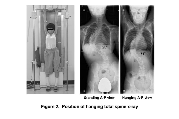
Background: Although most idiopathic scoliosis patients subject to conservative treatment in daily clinical practice, there have been no ideal methods to evaluate the spinal flexibility for the patients who are scheduled the brace treatment. The purpose of this study was to investigate the value of hanging total spine x-ray to estimate the indicative correction angle by brace wearing in idiopathic scoliosis patients.

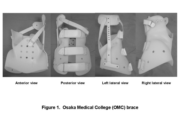
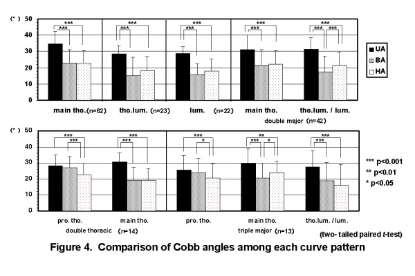
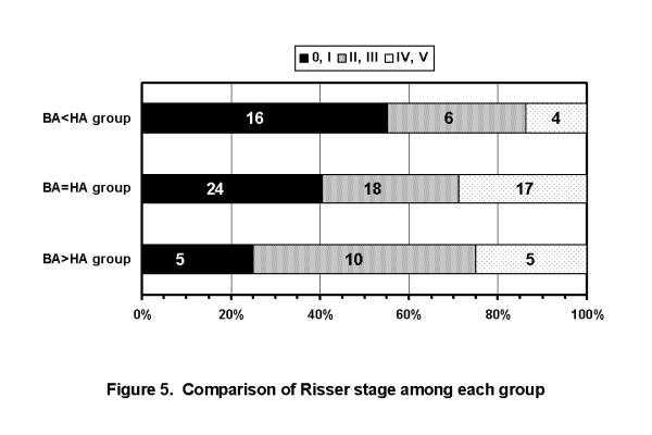
Methods: One hundred seventy-six consecutive patients with idiopathic scoliosis who were newly prescribed the Osaka Medical College (OMC) brace were studied. The study included 14 boys and 162 girls with a mean age of 13 years and 1 month. The type of curves consisted of 62 thoracic, 23 thoracolumbar, 22 lumbar, 42 double major, 14 double thoracic, and 13 triple curve pattern. We compared the Cobb angles on initial brace wearing (BA) and in hanging position (HA). Of those, 108 patients who had main thoracic curves were selected and evaluated the corrective ability of OMC brace. These subjects were divided into three groups according to the relation between BA and HA (BA < HA group, BA = HA group, and BA > HA group), and then, maturity was compared among them.

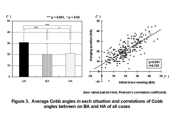
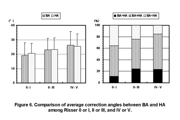
Results: The average Cobb angle in upright position (UA) of all cases was 31.0 ± 7.8°. The average BA and HA of all cases were 20.3 ± 9.5° and 21.1 ± 8.4°, respectively. The average chronological age was lowest in BA < HA group. And also, maturity in BA < HA group was the lowest among each of them. The rate of BA < HA cases were decreased as the Risser stage of the patients were progressed.

Conclusions: The use of hanging total spine x-ray served as a useful tool to estimate the degree of correction possible curve within the OMC brace for main thoracic curve in idiopathic scoliosis. Maturity had some influence on the correlation between HA and BA. Namely, in immature patients, HA tended to be larger than BA. In contrast, in mature patients, HA had a tendency to be smaller than BA. With consideration for spinal flexibility based on maturity, in mature patients, larger BA than HA may be allowed. However, in immature patients, smaller BA than HA should be aimed.


Figures

Figure 1
Figure 1
Osaka Medical College (OMC) brace. The OMC brace simply consists of a pelvic girdle, an upright bar, a high thoracic pad, and two straps. A pelvic girdle facilitates correction of a main thoracic curve. An upright bar with a high thoracic pad can correct a proximal thoracic curve utilizing righting reflex produced by patients' own response. Anterior and posterior straps prevent the body moving away from the brace during forward and backward bending motion.
Figure 2
Figure 2
Position of taking hanging total spine x-ray. Hanging total spine x-ray was taken in position that the patient is hanging onto the bar, stretching the back, and touching the toes lightly to the floor not to sway the body. Patients were instructed making a great effort to stretch their back as much as possible.
Figure 3
Figure 3
Average Cobb angles in each situation and correlation between on initial brace wearing and in hanging position of all cases. The average UA of all cases was 31.0 ± 7.8°. The average BA and HA of all cases were 20.3 ± 9.4° and 21.3 ± 8.4°, respectively. There were significant correlations between BA and HA. (UA: Cobb angle in upright position, BA: Cobb angle on initial brace wearing, HA: Cobb angle in hanging position).
Figure 4
Figure 4
Comparison of Cobb angles among each curve pattern. In single curve patterns, BA was almost the same as HA. In multiple curve patterns, BA of major curves was almost the same as HA of them, except thoracolumbar and lumbar curves in double major curve pattern, proximal thoracic curve in double thoracic curve pattern, and proximal and main thoracic curves in triple major curve pattern. (BA: Cobb angle on initial brace wearing, HA: Cobb angle in hanging position).
Figure 5
Figure 5
Comparison of Risser stage among each group. The rates of Risser 0 or I cases were 55.2% in BA < HA group, 40.7% in BA = HA group, and 25.0% in BA > HA group, respectively. Maturity in BA < HA group was the lowest among each of them. (BA: Cobb angle on initial brace wearing, HA: Cobb angle in hanging position).
Figure 6
Figure 6
Comparison of average Cobb angles between on initial brace wearing and in hanging position among Risser 0 or I, II or III, and IV or V. In the patients of Risser 0 or I, the average BA was smaller than the average HA. While, in the patients of Risser IV or V, the average HA was smaller than the average BA. The rate of BA < HA cases were decreased as the Risser stage of the patients were progressed. (BA: Cobb angle on initial brace wearing, HA: Cobb angle in hanging position).

Similar articles

Cited by

References

    1. Vaughan JJ, Winter RB, Lonstein JE. Comparison of the use of supine bending and traction radiographs in the selection of the fusion area in adolescent idiopathic scoliosis. Spine. 1996;21(21):2469–2473. doi: 10.1097/00007632-199611010-00012. - DOI - PubMed
    1. Kleinman RG, Csongradi JJ, Rinksy LA, Bleck EE. The radiographic assessment of spinal flexibility in scoliosis: a study of the efficacy of the prone push film. Clin Orthop. 1982;162:47–53. - PubMed
    1. Vedantam R, Lenke LG, Bridwell KH, Linville DL. Comparison of push-prone and lateral-bending radiographs for predicting postoperative coronal alignment in thoracolumbar and lumbar scoliotic curves. Spine. 2000;25(1):76–81. doi: 10.1097/00007632-200001010-00014. - DOI - PubMed
    1. Cheung KMC, Luk KDK. Prediction of correction of scoliosis with use of the fulcrum bending radiograph. J Bone Joint Surg. 1997;79A(8):1144–1150. - PubMed
    1. Luk KDK, Cheung KMC, Lu DS, Leong CY. Assessment of scoliosis correction in relation to flexibility using the fulcrum bending correction index. Spine. 1998;23(21):2303–2307. doi: 10.1097/00007632-199811010-00011. - DOI - PubMed

LinkOut - more resources