. 2012 Jun 21;119(25):5955-62.
doi: 10.1182/blood-2012-01-406801. Epub 2012 Mar 27.

Rational design and characterization of platelet factor 4 antagonists for the study of heparin-induced thrombocytopenia

Affiliations

Rational design and characterization of platelet factor 4 antagonists for the study of heparin-induced thrombocytopenia

Bruce S Sachais et al. Blood. .

Abstract

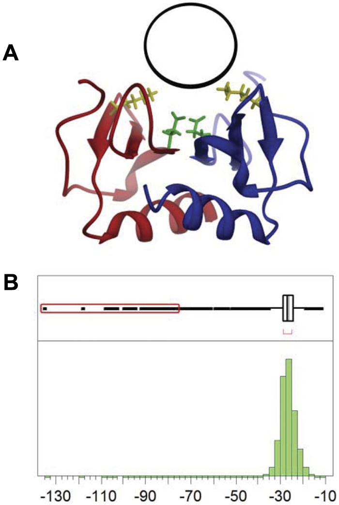
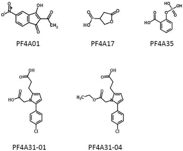
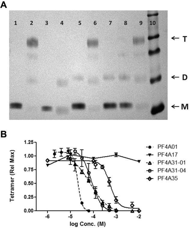
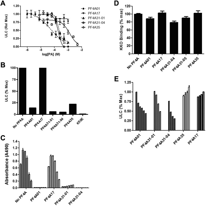
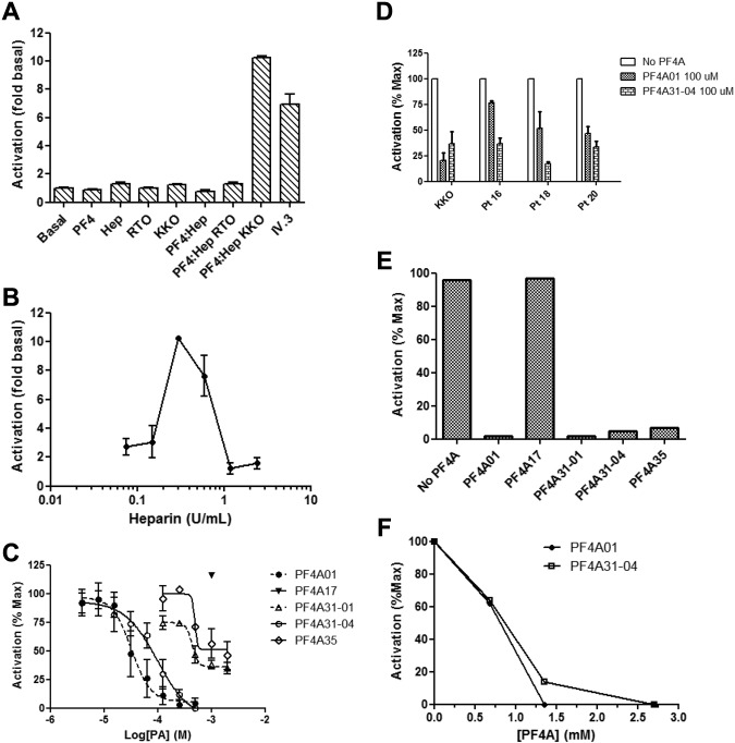
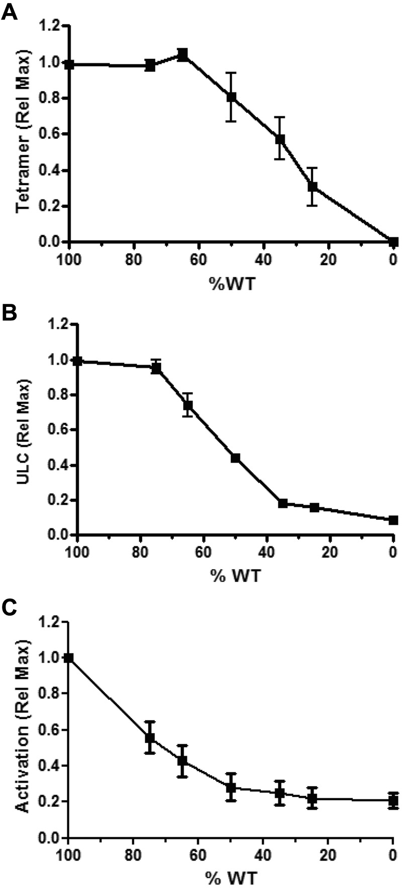
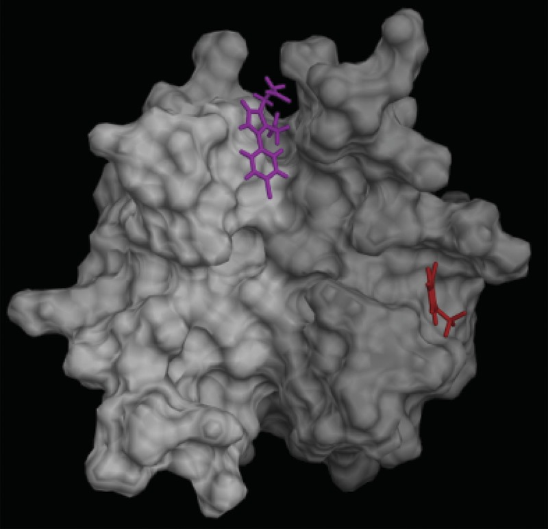
Patients with heparin-induced thrombocytopenia (HIT) remain at risk for recurrent thromboembolic complications despite improvements in management. HIT is caused by antibodies that preferentially recognize ultralarge complexes (ULCs) of heparin and platelet factor 4 (PF4) tetramers. We demonstrated previously that a variant PF4(K50E) forms dimers but does not tetramerize or form ULCs. Here, we identified small molecules predicted to bind PF4 near the dimer-dimer interface and that interfere with PF4 tetramerization. Screening a library of small molecules in silico for binding at this site, we identified 4 compounds that inhibited tetramerization at micromolar concentrations, designated PF4 antagonists (PF4As). PF4As also inhibited formation of pathogenic ULCs, and 3 of these PF4As promoted the breakdown of preformed ULCs. To characterize the ability of PF4As to inhibit cellular activation, we developed a robust and reproducible assay that measures cellular activation by HIT antibodies via FcγRIIA using DT40 cells. PF4As inhibit FcγRIIA-dependent activation of DT40 cells by HIT antibodies as well as platelet activation, as measured by serotonin release. PF4As provide new tools to probe the pathophysiology of HIT. They also may provide insight into the development of novel, disease-specific therapeutics for the treatment of thromboembolic complications in HIT.


Figures

Figure 1
Figure 1
Identification of binding site and computational screening. (A) The PF4 antagonist binding pocket (black circle) is located on the surface of the PF4 dimer (red and blue ribbons) near residues Lys50 (yellow) and Glu28 (green). (B) Distribution of DOCK scores for potential PF4 antagonists; 109 compounds have scores more than 10 SD below the mean (red box).
Figure 2
Figure 2
Structures of PF4 antagonists. Chemical structures of 5 compounds characterized in this article are shown.
Figure 3
Figure 3
Inhibition of PF4 tetramerization. (A) SDS-PAGE of PF4 cross-linked with BS3 (or uncross-linked controls) with or without antagonists (lane 1, WT-PF4; lane 2, WT-PF4 cross-linked; lane 3, PF4K50E; lane 4, PF4K50E cross-linked; lanes 5-9, WT-PF4 cross-linked in the presence of antagonist: lane 5, 250μM PF4A01; lane 6, 500μM PF4A17; lane 7, 250μM PF4A31-01; lane 8, 250μM PF4A31-04; lane 9, 500μM PF4A35; lane 10, molecular mass markers). Arrows denote PF4 monomers (M), dimers (D), and tetramers (T). (B) Dose-response inhibition of PF4 tetramer formation by antagonists. Data are the mean ± SEM of at least 3 independent experiments. Curves represent fit of data to the indirect Hill equation.
Figure 4
Figure 4
Inhibition of PF4:heparin ULCs. (A) Dose response of antagonists for inhibition of ULC formation measured by ELISA. Data are the mean ± SEM of at least 3 independent experiments performed in triplicate. Curves represent fit of data to the indirect Hill equation. (B) Inhibition of ULCs as measured by gel filtration. Antagonists are at 1mM. Data are representative of 2 or more experiments. (C) Inhibition of ULCs by PF4As spans a wide range of heparin concentrations. Conditions are as in panel A, except that ULCs were formed with PF4 and various concentrations of heparin (0.05, 0.1, 0.2, 0.4, 0.8, and 1.6 U/mL from left to right in each group). (D) Incubation of KKO with PF4As (1mM) does not inhibit KKO binding to ULCs as measured by ELISA. Data are the mean ± SEM of at least 3 independent experiments performed in triplicate. (E) Dose response of antagonists for breakdown of preformed ULCs. Data are mean ± SEM of at least 3 independent experiments performed in triplicate. Concentrations of antagonists for each series (from right to left) are 2mM, 1mM, 500μM, and 250μM (125μM and 63μM for PF4A01 only).
Figure 5
Figure 5
FcγRIIA-mediated activation of cells and inhibition by PF4 antagonists. (A) Activation of DT40 cells transfected with FcγRIIA and a luciferase reporter. Basal condition is a buffer only control, Hep indicates heparin, and IV.3 is the anti-FcγRIIA monoclonal antibody in the presence of anti-IgG antibody. (B) Dose response of DT40 activation by heparin in the presence of constant amounts of PF4. (C) Dose response of antagonists for inhibition of DT40 activation. Data are the mean ± SEM of at least 3 independent experiments performed in triplicate. Curves in panel C represent fit of data to the indirect Hill equation. (D) Inhibition of DT40 activation by plasma obtained from 3 HIT patients. Both antagonists completely inhibited activation by KKO and patient samples at a concentration of 500μM. Data represent the mean ± SEM of at least 2 independent experiments performed in triplicate. (E) Activation of platelets as measured by release of 14C-5-hydroxytryptamine creatinine sulfate. Data are representative of at least 2 experiments. Antagonists were present at a concentration of 2.5mM. (F) As in panel E, except that activation was by plasma from patients with HIT, and several antagonist concentrations were measured.
Figure 6
Figure 6
Inhibition of WT-PF4 with the nontetramerizing mutant PF4K50E. Inhibition of PF4 tetramerization (A), ULC formation (B), and FcγRIIA activation (C). PF4 concentrations were maintained constant by diluting WT PF4 with PF4K50E. Data are the mean ± SEM of at least 3 independent experiments performed in triplicate.
Figure 7
Figure 7
Predicted binding sites of PF4 antagonists. PF4 antagonists PF4A31 (magenta) and PF4A35 (red) are predicted to bind preferentially at different locations on the surface of the PF4 dimer (white-gray).

Comment in

References

    1. Arepally G, Cines DB. Heparin-induced thrombocytopenia and thrombosis. Clin Rev Allergy Immunol. 1998;16(3):237–247. - PubMed
    1. Warkentin TE. Heparin-induced thrombocytopenia: a ten-year retrospective. Annu Rev Med. 1999;50:129–147. - PubMed
    1. Arepally GM, Ortel TL. Heparin-induced thrombocytopenia. Annu Rev Med. 2010;61:77–90. - PMC - PubMed
    1. Amiral J, Bridey F, Dreyfus M, et al. Platelet factor 4 complexed to heparin is the target for antibodies generated in heparin-induced thrombocytopenia. Thromb Haemost. 1992;68(1):95–96. - PubMed
    1. Busch C, Dawes J, Pepper DS, Wasteson A. Binding of platelet factor 4 to cultured human umbilical vein endothelial cells. Thromb Res. 1980;19(1-2):129–137. - PubMed

Publication types

MeSH terms