. 2012 Mar 27:4:7.
doi: 10.1186/1758-3284-4-7.

Nimesulide inhibited the growth of hypopharyngeal carcinoma cells via suppressing Survivin expression

Affiliations

Nimesulide inhibited the growth of hypopharyngeal carcinoma cells via suppressing Survivin expression

Tian Jia-Jun et al. Head Neck Oncol. .

Expression of concern in

  • Comment: Head and Neck Oncology.
    BioMed Central. BioMed Central. BMC Med. 2014 Feb 5;12:24. doi: 10.1186/1741-7015-12-24. BMC Med. 2014. PMID: 24499430 Free PMC article. Review.

Abstract

Background: The objective of this study was to evaluate the efficacy of Nimesulide, a selective cyclooxygenase-2 (COX-2) inhibitor, on the growth of hypopharyngeal carcinoma cells (FaDu) in vitro, and investigate its potential mechanism.

Methods: After FaDu cells were treated with graded concentrations of Nimesulide for divergent time, sensitivity of cells to drug treatment was analyzed by MTT assay. Morphological changes of FaDu cells in the presence of Nimesulide were observed by acridine orange cytochemistry staining. Proliferating cells were detected using the 5-Bromo-2'-deoxy-uridine (BrdU) incorporation assay. Following cells were subjected to Nimesulide (500 μmol/l) for 6 h, 12 h and 24 h, the percentage of apoptosis was examined by flow cytometry. We detected COX-2 and Survivin expression change by RT-PCR and Western blot, and analyzed the correlation of them with the growth of FaDu cells. Additionally, we also analyzed Caspase-3, Bcl-2 and Bax expressions as markers to investigate the related pathway of Nimesulide-indued apoptosis.

Results: Compared with the control group, the viabilities rates were decreased by Nimesulide in time- and dose-dependent manners, typical morphological changes of apoptotic cells were observed in the Nimesulide-treatment groups, Nimesulide could suppress the proliferation of FaDu cells significantly. The percentage of apoptosis in FaDu cells were markedly increased after Nimesulide-treatment for 6 h, 12 h and 24 h. Nimesulide down-regulated the Survivin and COX-2 expressions at mRNA and protein levels in FaDu cells. Additional analyses indicated that Bcl-2 expression was significantly decreased and the expressions of Caspase-3 as well as Bax were increased at both mRNA and protein levels.

Conclusions: Based on the induction of apoptosis and suppression of proliferation, Nimesulide could inhibit the growth of FaDu cells. Furthermore, the suppression of Survivin expression may play an important role in Nimesulide-induced growth inhibition. Nimesulide could act as an effective therapeutic agent for hypopharyngeal carcinoma therapy.


Figures

Figure 1
Figure 1
Effects of Nimesulide on viability of FaDu cells. The anti-proliferating effects of Nimesulide were evaluated by MTT assay. Cells were treated with 0-1000 μM Nimesulide and the percentage of growth inhibition was determined after 12 ~ 48 h of treatment. FaDu cells had significant time- and dose-dependent growth inhibition due to Nimesulide.
Figure 2
Figure 2
Morphological changes of FaDu cells before and after Nimesulide-treatment (500 μmol/l, 24 h). Cells were stained with acridine orange and detected by fluorescence microscopy (×200). A: The number of survival cells in Nimesulide treated group was much fewer than that of control. B: Morphological changes and apoptotic bodies were detected obviously in Nimesulide group.
Figure 3
Figure 3
Inhibition of cell proliferation was detected by BrdU incorporation assay after treatment with Nimesulide (500 μmol/l). A: control group; B: treated with Nimesulide (500 μmol/l). a/d: The BrdU stained cells by immunofluorescence was detected by fluorescence microscopy (×200); b/e: nuclear stained by DAPI was detected by fluorescence microscopy (×200), c/f: Direct detected by reserved microscopy (×200).
Figure 4
Figure 4
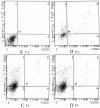
Apoptotic rates of FaDu cells treated with Nimesulide were detected using FCM. The percentage of apoptosis cells was markedly increased in the present of Nimesulide. × -axis represents the density of propidium iodide (PI), Y- axis represents the density of Annexin V-FITC. A: control; B: 6 h, C: 12 h; D: 24 h.
Figure 5
Figure 5
Alterations of Survivin and COX-2 expressions in FaDu cells. A: RT-PCR analysis. B: Western blot detection. The optical densities (OD) value of mRNA (C) and proteins (D) in each lane using NIH software Image J system were demonstrated in different times treated with Nimesulide. Each data point in the figure represents the mean ± SEM of three separate experiments.
Figure 6
Figure 6
The mRNA and protein levels of Bax, Bcl-2 and Caspase-3 in FaDu cells. A: RT-PCR analysis. B: Western blot measurement. The optical densities (OD) value of mRNA (C) and proteins (D) in each lane using NIH software Image J system were demonstrated in different times treated with Nimesulide. Each data point in the figure represents the mean ± SEM of three separate experiments.

References

    1. Tsujii M, DuBois RN. Alterations in cellular adhesion and apoptosis in epithelial cells overexpressing prostaglandin endoperoxide synthase 2. Cell. 1995;83(3):493–501. doi: 10.1016/0092-8674(95)90127-2. - DOI - PubMed
    1. Hu M, Peluffo G, Chen H, Gelman R, Schnitt S. et al.Role of COX-2 in epithelial-stromal cell interactions and progression of ductal carcinoma in situ of the breast. PNAS. 2009;106(9):3372–3377. doi: 10.1073/pnas.0813306106. - DOI - PMC - PubMed
    1. Ristimaki A, Sivula A, Lundin J, Lundin M, Salminen T. et al.Prognostic significance of elevated cyclooxygenase-2 expression in breast cancer. Cancer Res. 2002;62(3):632–635. - PubMed
    1. Zhang L, Tu J, Yu ZL, Wu YD, Xu CM. et al.Effects of the inhibition of cyclooxygenase-2 on human esophageal cancer cells: inhibition of cell proliferation and induction of apoptosis. Pathol Oncol Res. 2010;16(1):39–45. doi: 10.1007/s12253-009-9185-6. - DOI - PubMed
    1. Zhou Y, Ran J, Tang C, Wu J, Honghua L. et al.Effect of celecoxib on E-cadherin, VEGF, Microvessel density and apoptosis in gastric cancer. Cancer Biol Ther. 2007;6(2):269–275. doi: 10.4161/cbt.6.2.3629. - DOI - PubMed

Publication types

MeSH terms