. 2012 Aug;20(8):1489-500.
doi: 10.1038/mt.2012.44. Epub 2012 Mar 27.

Gene transfer corrects acute GM2 gangliosidosis--potential therapeutic contribution of perivascular enzyme flow

Affiliations

Gene transfer corrects acute GM2 gangliosidosis--potential therapeutic contribution of perivascular enzyme flow

M Begoña Cachón-González et al. Mol Ther. 2012 Aug.

Abstract

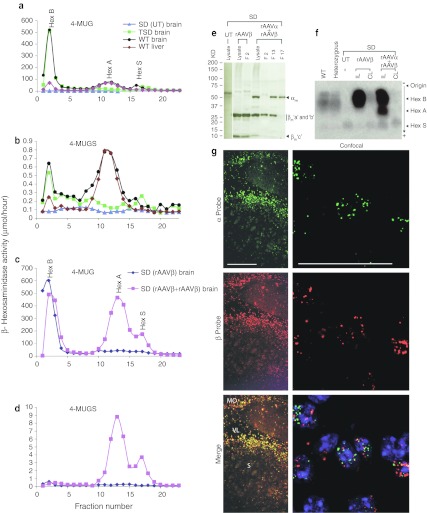
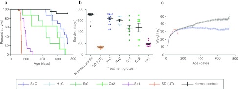
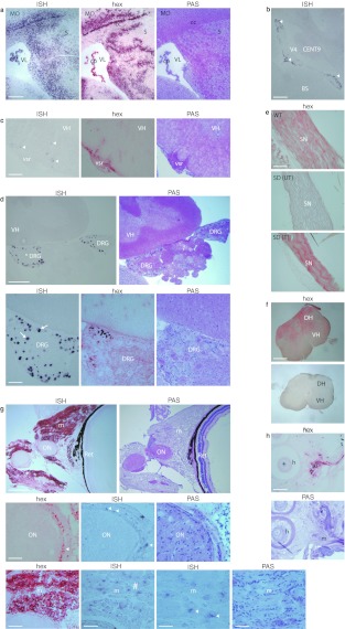
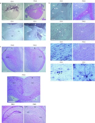
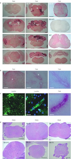
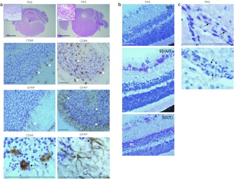
The GM2 gangliosidoses are fatal lysosomal storage diseases principally affecting the brain. Absence of β-hexosaminidase A and B activities in the Sandhoff mouse causes neurological dysfunction and recapitulates the acute Tay-Sachs (TSD) and Sandhoff diseases (SD) in infants. Intracranial coinjection of recombinant adeno-associated viral vectors (rAAV), serotype 2/1, expressing human β-hexosaminidase α (HEXA) and β (HEXB) subunits into 1-month-old Sandhoff mice gave unprecedented survival to 2 years and prevented disease throughout the brain and spinal cord. Classical manifestations of disease, including spasticity-as opposed to tremor-ataxia-were resolved by localized gene transfer to the striatum or cerebellum, respectively. Abundant biosynthesis of β-hexosaminidase isozymes and their global distribution via axonal, perivascular, and cerebrospinal fluid (CSF) spaces, as well as diffusion, account for the sustained phenotypic rescue-long-term protein expression by transduced brain parenchyma, choroid plexus epithelium, and dorsal root ganglia neurons supplies the corrective enzyme. Prolonged survival permitted expression of cryptic disease in organs not accessed by intracranial vector delivery. We contend that infusion of rAAV into CSF space and intraparenchymal administration by convection-enhanced delivery at a few strategic sites will optimally treat neurodegeneration in many diseases affecting the nervous system.


Figures

Figure 1
Figure 1
β-Hexosaminidase profile of mouse tissues. (ad) Representative activities of β-hexosaminidase isozymes Hex A, Hex B, and Hex S in liver or brain extracts separated by ion-exchange chromatography; wild type (WT), untreated (UT), Sandhoff (SD), and Tay–Sachs (TSD) disease mice; rAAVβ or rAAVα+rAAVβ transduced brain was analyzed 4 weeks postinjection. Activities of 5 µl samples were determined against substrates 4-MUG and 4-MUGS, and normalized to 100 µg of loaded protein. (e) β-Hexosaminidase polypeptide components resolved by sodium dodecyl sulfate-polyacrylamide gel electrophoresis (SDS-PAGE), and immunoblotted with goat antihuman Hex A: unfractionated lysate (lysate) and fractions 2 (F 2), 13 (F 13) and 17 (F 17) in panels c and d. αm, mature α subunit; βm”a,” βm”b”, and βm”c,” proteolysed species of mature β subunit. Standards are given in kDa. (f) Cellulose-acetate electrophoresis of extracts from WT, hexb heterozygous, and SD mouse cerebrum injected at a single striatal site. Isozymes from ipsilateral (IL) and contralateral (CL) hemispheres revealed by incubation with 4-MUG. Micrograms of total protein loaded (from left to right): 103, 108, 242, 120, 128, 185, and 229. (g) Human α and β subunits RNA detected in SD (rAAVα+rAAVβ) brain sections by in situ hybridization with fluorescein and cyanine-3-conjugated tyramide signal amplification reagent, respectively. Mice were injected at one month and killed aged 2 years. Diamidino-2-phenylindole (DAPI)-stains cell nuclei blue. Bars: 200 µm (left panels in g); 50 µm (right panels in g). rAAV, recombinant adeno-associated viral vectors.
Figure 2
Figure 2
Lifespan of Sandhoff mice after intracranial infusions of rAAVα+rAAVβ at 1 month of age. (a) Kaplan–Meier survival curve, data censored at 2 years. Sandhoff (SD) mice were injected unilaterally into the striatum (Sx1, n = 15), bilaterally into the striatum (Sx2, n = 8) or cerebellum (Cx2, n = 8), and bilaterally into the striatum and cerebellum (S+C, n = 16) or into the hippocampus and cerebellum (H+C, n = 8). Control groups: untreated normal controls (n = 22) and SD mice [SD (UT), n = 37]. (b) One-way analysis of variance (ANOVA) and Bonferroni multiple post-hoc comparisons with mean ± SEM. (c) Comparison of average weekly weights for S+C-treated, normal controls and SD (UT) groups. The mean ± 1.96 SE are plotted against the mid-point of each week. rAAV, recombinant adeno-associated viral vectors.
Figure 3
Figure 3
Biodistribution of viral vector and enzyme. (ad) Detection of vector RNA on sections from Sandhoff (SD) mice injected with rAAVα+rAAVβ into the striatum at one month of age by in situ hybridization (ISH, black stain). ISH stained: gray and white matter, such as (a) corpus callosum (cc); choroid plexus (cp) in all ventricles, including that in (a) lateral ventricles (VL), and (b) 4th ventricle (V4) (arrowheads); ventral roots of the (c) spinal cord (vsr) (arrowheads); and (d) dorsal root ganglia (DRG) (arrows in magnified view of asterisk region). Consecutive sections stained for enzyme (hex), red precipitate, positively correlated with ISH-labeled regions, as shown in panels a, c, and d. Enzyme was present in (e) sciatic nerve; and (f) spinal cord and roots (top panel treated, bottom panel untreated SD). Vector RNA and enzyme was found extracranially in the orbit, around optic nerve (ON) (arrowheads) and periocular muscle (m) (arrows in magnification of area with hash symbol) (g); and (h) subcutaneous muscle supporting hair follicles. Consecutive sections stained with periodic acid-Schiff (PAS) for structure identification. Wild type (WT), untreated [SD (UT)] and rAAVα+rAAVβ-treated [SD (T)] Sandhoff mice. Brain stem (BS); dorsal horn (DH); motor cortex (MO); ninth cerebellar lobule (CENT9); retina (Ret); striatum (S); ventral horn (VH). Bars: 500 µm (a, f, top panels in d and g); 200 µm (e, h, and lower panels in d); 100 µm (b, c; and first and second left lower panels in g); 50 µm (middle panels in g; and third and fourth right lower panels in g). rAAV, recombinant adeno-associated viral vectors.
Figure 4
Figure 4
Resolution of pathology in brain regions distant from injected sites. (a) In situ hybridization (ISH) staining, black precipitate, for vector RNA of sections from a Cx2-injected SD mouse was heaviest in right cerebellum and dorsal brain stem nuclei (BS) (top left panel and magnified views of hash and asterisk areas in lower panels). (b) ISH stained Purkinje cell layer (PKL), and pontine nucleus (PN) (arrowheads). Cells in white matter, such as middle cerebellar peduncle (mcp), had oligodendrocytic (black arrows) or other glial (white arrow) morphologies. (c) In this animal, injected at 1 month and killed at 628 days of age (Supplementary Video S7, viewed at 1 year), glycoconjugate accumulation, magenta stained with periodic acid-Schiff (PAS) reagent, was principally found in left brain; shown are olfactory bulbs (top panels), motor cortex (MO) (middle panels), and hippocampus (lower panels). Anterior olfactory nucleus, medial part (AOM); field CA1 and CA3 of hippocampus; corpus callosum (cc); dentate gyrus (DG); fourth ventricle (V4); lateral ventricle (VL); molecular (ML) and granular (GL) layers of the cerebellar cortex; myelin tracts (my); right (R) and left (L) hemispheres. Bars: 2 mm (top panels in a); 500 µm (c and lower panels in a); 200 µm (from top: first and second set of panels in b); 100 µm (from top: third and fourth set of panels in b).
Figure 5
Figure 5
β-Hexosaminidase with prominent perivascular localization reduces glycoconjugate storage throughout the neuraxis. (a) In a representative S+C-injected Sandhoff mouse [SD (T)] β-hexosaminidase stain (hex), red precipitate, was found throughout the brain (shown from top left to middle lower panel: forebrain through to hindbrain), and spinal cord (top rightmost panel); viewed at 1 and 2 years of age in Supplementary Videos S2 and S3. Controls: 4-month untreated Sandhoff [SD (UT)] (Supplementary Video S1) and 2-year-old wild type (WT). (b) Much of the enzyme stain appears perivascular; magnified views: area of asterisk in a (left panel), cross (middle panel) and hash (right panel) areas in b. (c) Costaining of hex with fluorescein-conjugated tomato lectin to label endothelium, showed close association—with hex also distributed away from the fluorescein label seen as a dark halo (left and middle panels). Diamidino-2-phenylindole (DAPI) stains cell nuclei blue. (d) Glycoconjugate material (magenta), stained with periodic acid-Schiff (PAS) reagent, at different levels of the spinal cord. Bars: 2 mm (brain panels in a); 500 µm (spinal cord panels in a and d; and left panel in b); 200 µm (left panel in c); 100 µm (middle panel in b); 50 µm (right panel in b, middle and right panels in c).
Figure 6
Figure 6
Inflammatory response in the presence of residual storage and emerging disease in nontargeted tissues. (a) The intensity and number of cells staining for the microglial marker, CD68, and astrocytic marker, GFAP, correlated with local glycoconjugate abundance (stained magenta with periodic acid-Schiff (PAS) reagent). Displayed are sections from an Sx2-injected Sandhoff (SD) mouse, killed at 696 days and viewed in Supplementary Videos S4 and S5 at 12 and 20 months of age. The ninth cerebellar lobule with few PAS-stained neurons (hash area and inset in left panel) had few cells labeled with CD68 (arrowheads in left panel) and GFAP (arrowhead in left panel). In contrast, the flocculus lobe with many intensely PAS-stained neurons (asterisk area and inset in right panel) had many cells labeled with CD68 (arrowheads, right panel) and GFAP (arrowheads, right panel). Globular microglia (arrows in left bottom panel) and astrocytes (arrows, right bottom panel) are intensely stained. (b) Abundant periodic acid-Schiff (PAS)-stained glycoconjugate was found in retinal ganglion cell layer (GL) in 4-month untreated SD [SD (UT)] compared with 2-year-old wild type (WT). In S+C-treated 2 year-old SD [SD (T)] staining was found in GL and inner nuclear (INL) layers. (c) SD (T) mouse endothelium in regions with low activity of β-hexosaminidase contained PAS-stained glycoconjugates (arrows, top panel), but staining was undetectable in regions with abundant enzyme activity such as those close to injection sites (arrows in lower panel). Bars: 2 mm (from top: first set of panels in a); 200 µm (insets in a); 100 µm (c, and from top: second, third, and fourth sets of panels in a); 50 µm (b).

References

    1. Johnson WG. The clinical spectrum of hexosaminidase deficiency diseases. Neurology. 1981;31:1453–1456. - PubMed
    1. Kolodny EH, Brady RO., and, Volk BW. Demonstration of an alteration of ganglioside metabolism in Tay-Sachs disease. Biochem Biophys Res Commun. 1969;37:526–531. - PubMed
    1. Okada S., and, O'Brien JS. Tay-Sachs disease: generalized absence of a beta-D-N-acetylhexosaminidase component. Science. 1969;165:698–700. - PubMed
    1. Sandhoff K. Variation of beta-N-acetylhexosaminidase-pattern in Tay-Sachs disease. FEBS Lett. 1969;4:351–354. - PubMed
    1. Conzelmann E., and, Sandhoff K. Purification and characterization of an activator protein for the degradation of glycolipids GM2 and GA2 by hexosaminidase A. Hoppe-Seyler's Z Physiol Chem. 1979;360:1837–1849. - PubMed

Publication types