. 2012 May 18;287(21):17789-17800.
doi: 10.1074/jbc.M111.304782. Epub 2012 Mar 27.

The FF4 and FF5 domains of transcription elongation regulator 1 (TCERG1) target proteins to the periphery of speckles

Affiliations

The FF4 and FF5 domains of transcription elongation regulator 1 (TCERG1) target proteins to the periphery of speckles

Noemí Sánchez-Hernández et al. J Biol Chem. .

Abstract

Transcription elongation regulator 1 (TCERG1) is a human factor implicated in interactions with the spliceosome as a coupler of transcription and splicing. The protein is highly concentrated at the interface between speckles (the compartments enriched in splicing factors) and nearby transcription sites. Here, we identified the FF4 and FF5 domains of TCERG1 as the amino acid sequences required to direct this protein to the periphery of nuclear speckles, where coordinated transcription/RNA processing events occur. Consistent with our localization data, we observed that the FF4 and FF5 pair is required to fold in solution, thus suggesting that the pair forms a functional unit. When added to heterologous proteins, the FF4-FF5 pair is capable of targeting the resulting fusion protein to speckles. This represents, to our knowledge, the first description of a targeting signal for the localization of proteins to sites peripheral to speckled domains. Moreover, this "speckle periphery-targeting signal" contributes to the regulation of alternative splicing decisions of a reporter pre-mRNA in vivo.


Figures

FIGURE 1.
FIGURE 1.
The FF4/FF5 domains are required for efficient targeting of TCERG1 to the speckle compartment. A, dual-labeling of cells transfected with ECFP-TCERG1[1–1098] or the indicated mutant constructs (ECFP, green) with the anti-SC35 antibody (SC35, red) was performed. A diagrammatic representation of the ECFP-TCERG1 fusion proteins is shown to the left of each panel. The numbers in parentheses represent the TCERG1 amino acids contained in the construct. Shown are the three WW domains, the putative nuclear localization signal (NLS), and the six FF domains. Line scans showing local intensity distributions of TCERG1 and SC35 are shown to the right of the panels. Bars in the merged panels indicate the positions of the line scans. Scale bars, 3 μm. B, the nucleoplasmic fraction of TCERG1 proteins present in speckles was determined after measuring the fluorescence intensity in speckles relative to nucleoplasm as described under “Experimental Procedures.” The data shown are from three independent experiments. Student's t test was performed, and differences were shown to be highly significant: p(TCERG1[1–1098] versus TCERG1[1–662]) < 0.0001; p(TCERG1[1–1098] versus TCERG1[1–662]-FF4) = 0.0001; p(TCERG1[1–1098] versus TCERG1[1–662]-FF5) = 0.0011; p(TCERG1[1–1098] versus TCERG1[1–662]-FF4/FF5) = 0.8261; p(TCERG1[1–1098] versus TCERG1[1–662]-FF5/FF6) = 0.0023; p(TCERG1[1–662]-FF4/FF5 versus TCERG1[1–662]-FF4) = 0.0060; p(TCERG1[1–662]-FF4/FF5 versus TCERG1[1–662]-FF5) = 0.0106; and p(TCERG1[1–662]-FF4/FF5 versus TCERG1[1–662]-FF5/FF6) = 0.0192. The bar graph represents mean ± S.D. C, immunofluorescence analysis of cells transfected with the indicated plasmids. Images show a speckled pattern for full-length TCERG1[1–1098], but TCERG1[1–662], TCERG1[ΔFF4/FF5], and TCERG1[ΔFF5] are more diffusely dispersed throughout the nucleoplasm. Individual and merged images of the cell labeled with the indicated fluorescent proteins (FP, in green) and with the anti-SC35 antibody (SC35, red) are shown. Scale bars = 3 μm.
FIGURE 2.
FIGURE 2.
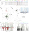
15N-HSQC NMR spectra and thermal unfolding of FF4-FF5. A, amino acid alignment of TCERG1 homologues. Secondary structure elements of FF4 and FF5 are drawn above the sequences in green and orange, respectively. Boxed sequences in blue are the residues that form the α-helices. The overlapped residues between FF4 and FF5 are shown in a red box. Strictly conserved residues are highlighted in red. B, 15N-HSQC spectra at 298 K for FF5 and FF4-FF5 constructs. The NMR data show the improvement of FF5 backbone amino acid dispersion (shown in black) observed in the pair when compared with that of the isolated construct. C, overlapped 15N-HSQC spectra at 298 K for FF4 and FF4-FF5 constructs and chemical shift differences bar representation of FF4 residues. D, thermal unfolding of the FF4 and FF4-FF5 domains as monitored by DSC. The difference in Tm is shown as an arrow connecting both maxima. E, secondary structure elements of the FF4-FF5 pair. Chemical shift distribution of Cα and Cβ. The ratio of Cα and Cβ is shown as a green line. The majorities of the values are positive, indicating the presence of a helical structure. F, heteronuclear {1H}-15N NOE. Unassigned residues, proline residues that lack a proton amide, and overlapped peaks were excluded from the analysis. Heteronuclear NOE values show that the pair of FF4-FF5 has secondary structure throughout the construct.
FIGURE 3.
FIGURE 3.
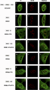
FF4/FF5 directs SRSF1 domain-deletion mutants to nuclear speckles. Cells were transfected with the indicated plasmids and dually labeled with antibodies directed against the expressed SRSF1 protein (left column, green) and SC35 (center column, red). The merged images are also shown (right column). In all cases, colocalization of expressed proteins with the endogenous marker was assessed by confocal imaging. A diagrammatic representation of the T7-tagged SRSF1 mutants used is shown at the left of the figure. The structure of the SRSF1 domain-deletion mutants was described previously (51). Scale bars = 3 μm.
FIGURE 4.
FIGURE 4.
Colocalization of the FF4/FF5-containing RRM1 protein with nuclear speckles is not perturbed following inhibition of transcription. HeLa cells were transfected with the indicated expression plasmids and treated with 25 μg/ml of α-amanitin for 6 h at 37 °C and then processed for immunofluorescence analysis. Dual-labeling of cells with antibodies directed against SRSF1 (T7, green) and with the SC35 antibody (red) was performed. Individual staining and merge images of the cell stained with the indicated antibodies are shown. A diagrammatic representation of the T7-tagged SRSF1 mutants used is shown at the left of the figure. Scale bars = 3 μm.
FIGURE 5.
FIGURE 5.
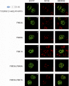
Strictly conserved phenylalanine residues within the FF4/FF5 domains are required for the targeting to nuclear speckles. Dual labeling of HEK293T cells transfected with TCERG1[1–662]-FF4/FF5 or the indicated phenylalanine-to-alanine mutant constructs (ECFP, green) with the anti-SC35 antibody (SC35, red) was performed. Individual and merged images of the cell are shown. Scale bars = 3 μm.
FIGURE 6.
FIGURE 6.
Deletion of the FF4/FF5 domains of TCERG1 affects Bcl-x alternative splicing. A, schematic representation of the structure of the Bcl-x minigene is drawn with exons (boxes) and introns (lines). The Bcl-x pre-mRNA is alternatively spliced (dotted lines) to produce two major isoforms, Bcl-xL and Bcl-xS. B, splicing assay of the Bcl-x minigene in HEK293T cells transfected with the indicated constructs. Cells were harvested ∼44 h after transfection and processed for RT-PCR. The data are presented as the ratio of Bcl-xL to Bcl-xS from three independent experiments (mean ± S.D.). *, p < 0.05.

Similar articles

Cited by

References

    1. Lamond A. I., Spector D. L. (2003) Nuclear speckles. A model for nuclear organelles. Nat. Rev. Mol. Cell Biol. 4, 605–612 - PubMed
    1. Spector D. L. (1990) Higher order nuclear organization. Three-dimensional distribution of small nuclear ribonucleoprotein particles. Proc. Natl. Acad. Sci. U.S.A. 87, 147–151 - PMC - PubMed
    1. Spector D. L., Fu X. D., Maniatis T. (1991) Associations between distinct pre-mRNA splicing components and the cell nucleus. EMBO J. 10, 3467–3481 - PMC - PubMed
    1. Huang S., Spector D. L. (1992) U1 and U2 small nuclear RNAs are present in nuclear speckles. Proc. Natl. Acad. Sci. U.S.A. 89, 305–308 - PMC - PubMed
    1. Fu X. D., Maniatis T. (1990) Factor required for mammalian spliceosome assembly is localized to discrete regions in the nucleus. Nature 343, 437–441 - PubMed

Publication types

MeSH terms

Substances

LinkOut - more resources