. 2012 May 11;287(20):16199-208.
doi: 10.1074/jbc.M111.335216. Epub 2012 Mar 27.

Kaposi sarcoma-associated herpesvirus vIRF-3 protein binds to F-box of Skp2 protein and acts as a regulator of c-Myc protein function and stability

Affiliations

Kaposi sarcoma-associated herpesvirus vIRF-3 protein binds to F-box of Skp2 protein and acts as a regulator of c-Myc protein function and stability

Petra Baresova et al. J Biol Chem. .

Abstract

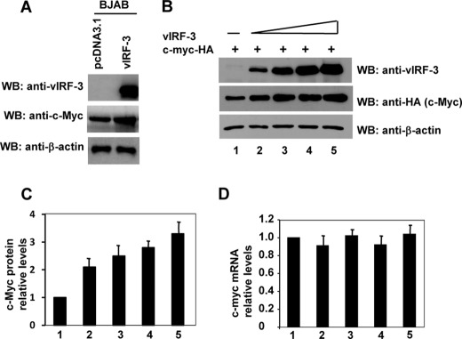
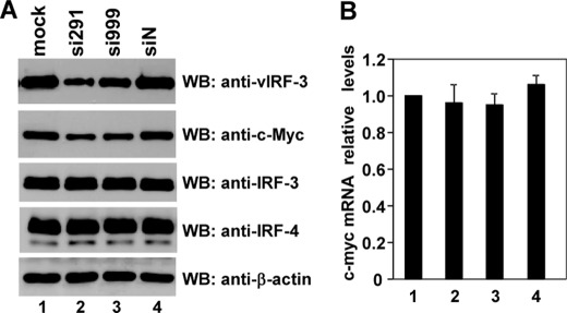
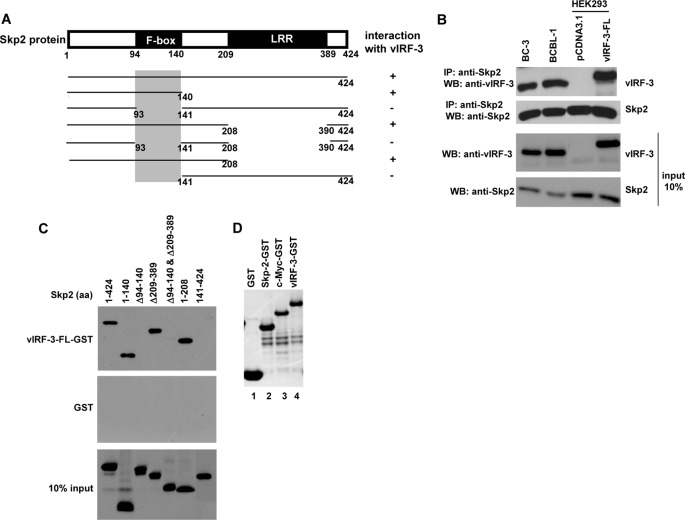
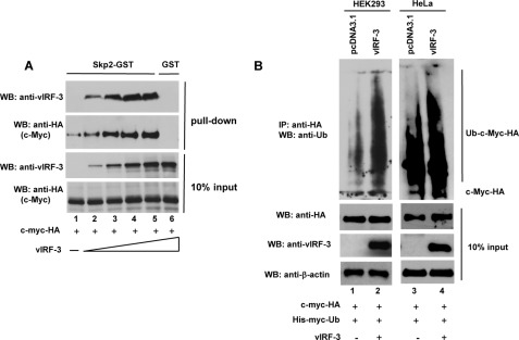
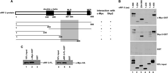
The Kaposi sarcoma-associated herpesvirus (KSHV) has been linked to Kaposi sarcoma, body cavity-based lymphoma, and Castleman disease. vIRF-3 is a KSHV latent gene that is critical for proliferation of KSHV-positive lymphoid cells. Furthermore, vIRF-3 contributes to KSHV-associated pathogenesis by stimulating c-Myc transcription activity. Here we show that vIRF-3 can associate with Skp2, a key component of the SCF(skp2) ubiquitin ligase complex. Skp2 is a transcriptional co-factor for c-Myc that was shown to regulate the stability of c-Myc protein as well as c-Myc-dependent transcription. In this study, we show that vIRF-3 binds to the F-box of Skp2 and recruits it to c-Myc-regulated promoters to activate c-Myc-dependent transcription. Additionally, cells overexpressing vIRF-3 exhibit higher levels of c-Myc ubiquitylation, suggesting that ubiquitylation is necessary for c-Myc-mediated transcription. Moreover, vIRF-3 can stabilize the c-Myc protein by increasing its half-life. Collectively, these results indicate that vIRF-3 can effectively manipulate c-Myc stability and function and thus contribute to c-Myc-induced KSHV-associated lymphomagenesis.


Figures

FIGURE 1.
FIGURE 1.
c-Myc stability is enhanced in presence of vIRF-3. A, BJAB stable cell lines expressing vIRF-3 (BJAB/vIRF-3) show higher levels of c-Myc protein when compared with control cells (BJAB/pcDNA3.1). WB, Western blot. B, HeLa cells were co-transfected with constant amounts of c-myc-HA and increasing amounts of vIRF-3 expression constructs (0, 0.5, 1.5, 3.0, and 6.0 μg). Forty-eight hours after transfection, cell extracts were prepared. The proteins in the extracts were separated on a 10% polyacrylamide gel and blotted with anti-HA (c-Myc), anti-vIRF-3, and anti-β-actin antibodies. C, quantitation of the immunoblot signals from panel B. c-Myc was normalized to the β-actin signal and then plotted against the signal obtained at 0 μg of vIRF-3 transfection. D, total RNA was isolated from cells used in panel B and analyzed by real-time RT-PCR using c-myc- and gapdh-specific probes. Error bars represent standard errors for four independent experiments. Each experiment was performed in duplicates.
FIGURE 2.
FIGURE 2.
Reduction of c-Myc protein in PEL cells after transient knockdown of vIRF-3 expression. A, BC-3 cells were either mock-treated or transfected with nonsense siRNA (siN) or vIRF-3-specific siRNAs (si291 and si999). At 2 days after transfection, half of the cells were harvested, and 8 μg of protein was analyzed by Western blot (WB) using anti-vIRF-3, anti-c-Myc, anti-β-actin, anti-IRF-3, and anti-IRF-4 antibodies. B, the other half of the cells used in panel A was harvested for total RNA isolation and analyzed by real-time RT-PCR using c-myc- and gapdh-specific probes. Error bars represent standard errors for four independent experiments. Each experiment was performed in duplicates.
FIGURE 3.
FIGURE 3.
Expression of vIRF-3 extends half-life of c-Myc. A, HeLa cells were co-transfected with constant amounts of c-myc-HA, vIRF-3, or pcDNA3.1 expression constructs. Forty-eight hours after transfection, cells were treated with 25 μg/ml cycloheximide (CHX) for the indicated times. Cell lysates were analyzed by Western blotting using anti-HA (c-Myc), anti-vIRF-3, and anti-β-actin antibodies. B, quantitation of the immunoblot signals from panel A. c-Myc was normalized to the β-actin signal and then plotted against the signal obtained at 0 h of cycloheximide treatment. Error bars represent standard errors for four independent experiments. Each experiment was performed in duplicates.
FIGURE 4.
FIGURE 4.
vIRF-3 interacts with F-box of Skp2. A, schematic representation of Skp2 deletion mutants tagged with Myc tag. The shaded box indicates the vIRF-3-binding domain. LRR, leucine-rich region. B, co-immunoprecipitation of Skp2 and vIRF-3 in PEL cells, BC-3 and BCBL-1, and transfected HEK293 cells. Protein lysates (400 μg) were immunoprecipitated (IP) with anti-Skp2 antibodies, and the immunoprecipitated complexes were analyzed by Western blot (WB) with anti-vIRF-3 antibodies. The relative levels of Skp2 and vIRF-3 in 40 μg of protein lysates are shown for comparison (input 10 %). The higher molecular weight of transfected vIRF-3-FL is due to Myc-His tag at the carboxyl terminus. C, Myc-tagged skp2 deletion mutants were in vitro translated using the TnT T7 quick coupled transcription/translation system and incubated with vIRF-3-FL fused to GST or GST alone immobilized on glutathione-Sepharose beads. The bound proteins were eluted and resolved on 10% SDS-PAGE followed by Western blot with anti-Myc (9E10) antibodies. 10% of Skp2 deletion mutant protein input is shown below (10% input). aa, amino acids. D, Coomassie Blue staining of purified GST, Skp2-GST, c-Myc-GST, and vIRF-3-GST used in GST pulldown experiments.
FIGURE 5.
FIGURE 5.
Analysis of c-Myc interaction with vIRF-3. A, schematic representation of c-myc deletion constructs. The shaded boxes indicate vIRF-3-binding domains. MBI and MBII, Myc conserved domains I and II; TAD, transcription activation domain; NLS, nuclear localization signal; BR, basic region; HLH, helix-loop-helix; LZ, leucine zipper. B, HA-tagged c-Myc deletion mutants were in vitro translated using the TnT T7 quick coupled transcription/translation system and incubated with vIRF-3-FL fused to GST or GST alone immobilized on glutathione-Sepharose beads. The bound proteins were eluted and resolved on 10% SDS-PAGE followed by Western blot with anti-HA antibodies. 10% of c-Myc deletion mutant protein input is shown below (10% input). aa, amino acids; N.S., nonspecific.
FIGURE 6.
FIGURE 6.
vIRF-3 neither inhibits c-Myc/Skp2 interaction nor blocks c-Myc ubiquitylation. A, increased binding of c-Myc to Skp2 in the presence of vIRF-3. Constant amounts of in vitro translated c-Myc-HA protein were incubated with Skp2-GST immobilized to glutathione-Sepharose beads in the presence of increasing amounts of in vitro translated vIRF-3. The bound proteins were eluted and resolved on 10% SDS-PAGE followed by Western blot (WB) with anti-HA (c-Myc) and anti-vIRF-3 antibodies. The binding to GST beads represents negative control (lane 6). 10% of c-Myc and vIRF-3 protein input is shown below (10% input). B, increased ubiquitylation of c-Myc in vIRF-3-expressing cells. HEK293 (lanes 1 and 2) and HeLa (lanes 3 and 4) cells were co-transfected with equal amounts of c-Myc-HA and His-Myc-Ub followed by transfection with either an empty vector (pcDNA3.1) or the vIRF-3-expressing construct. Prior to lysis, the cells were pretreated with proteasome inhibitor, 50 μm MG115, for 3 h. The cell lysates (400 μg) were analyzed by immunoprecipitation (IP) with anti-HA-specific antibodies followed by Western blotting with anti-Ub antibodies. The relative levels of transfected c-Myc-HA and vIRF-3 in 40 μg of cell lysates were estimated by Western blots (10% input).
FIGURE 7.
FIGURE 7.
vIRF-3 cooperates with Skp-2 in activation of c-Myc-mediated transcription. A, effect of vIRF-3 on activation of cdk4 promoter. Human wild-type cdk4 reporter (WT MBS1–4) and pRL-SV40 plasmids were co-transfected into HEK293 cells with either an empty vector (pcDNA3.1) or c-myc-HA-, vIRF-3-, and skp2-Myc-expressing plasmids. Luciferase activity was analyzed 48 h after transfection as -fold activation relative to the basal level of reporter gene in the presence of control vector, pcDNA3.1. Results were normalized to Renilla luciferase activity. Error bars represent standard errors for three independent experiments. The bottom panel shows the expression levels of transfected proteins. RLU, relative luciferase units. B, association of c-Myc, vIRF-3, and Skp2 with the WT cdk4 promoter was analyzed by DNA pulldown assay. HEK293 cells were transfected with c-myc-HA-, vIRF-3-, and skp2-Myc-expressing plasmids as indicated. Twenty-four hours after transfection, cells were untreated or treated with 50 μm MG115 for 3 h followed by cell lysis. Lysates (350 μg) were incubated with WT cdk4 promoter (WT MBS4) oligodeoxynucleotides coupled to magnetic beads. The c-Myc-HA, vIRF-3, or Skp2-Myc proteins pulled down by DNA were identified by Western blot with anti-HA, anti-vIRF-3, and anti-Myc (9E10)-specific antibodies, respectively (upper panel). Binding to beads only (lanes 3 and 6) represents the negative control. Binding to mutated cdk4 promoter (mutMBS4) served as a negative control (middle panel). The relative levels of transfected vIRF-3, c-Myc-HA, and Skp-2-Myc in 35 μg of cell extracts were estimated by Western blot (10% input, bottom panel).
FIGURE 8.
FIGURE 8.
c-Myc and Skp2 interact with vIRF-3 (amino acids 346–455). A, schematic representation of vIRF-3 N- and C-terminal deletion mutants. The shaded box indicates the common domain for c-Myc and Skp2 binding. NLS, nuclear localization signal; NES, nuclear export signal. B, vIRF-3 deletion mutants were in vitro translated using TnT T7 quick coupled transcription/translation system and incubated with c-Myc or Skp2 fused to GST or GST alone immobilized on glutathione-Sepharose beads. The bound proteins were eluted and resolved on 10% SDS-PAGE followed by Western blot with anti-vIRF-3 antibodies. 10% of vIRF-3 deletion mutant protein input is shown below (10% input). aa, amino acids. C, vIRF-3 (left panel) or c-Myc-HA (right panel) was in vitro translated using E. coli T7 S30 extract system and incubated with c-Myc-GST or vIRF-3-FL-GST, respectively. Incubation with GST alone served as a negative control. The bound proteins were eluted and resolved on 10% SDS-PAGE followed by Western blot with either anti-vIRF-3 or anti-HA antibodies. 10% of in vitro translated vIRF-3 or c-Myc-HA is also shown (10% input).

Similar articles

Cited by

References

    1. Moore P. S., Gao S. J., Dominguez G., Cesarman E., Lungu O., Knowles D. M., Garber R., Pellett P. E., McGeoch D. J., Chang Y. (1996) Primary characterization of a herpesvirus agent associated with Kaposi's sarcomae. J. Virol. 70, 549–558 - PMC - PubMed
    1. Lee H. R., Kim M. H., Lee J. S., Liang C., Jung J. U. (2009) Viral interferon regulatory factors. J. Interferon Cytokine Res. 29, 621–627 - PMC - PubMed
    1. Lubyova B., Pitha P. M. (2000) Characterization of a novel human herpesvirus 8-encoded protein, vIRF-3, that shows homology to viral and cellular interferon regulatory factors. J. Virol. 74, 8194–8201 - PMC - PubMed
    1. Rivas C., Thlick A. E., Parravicini C., Moore P. S., Chang Y. (2001) Kaposi sarcoma-associated herpesvirus LANA2 is a B-cell-specific latent viral protein that inhibits p53. J. Virol. 75, 429–438 - PMC - PubMed
    1. Cesarman E., Chang Y., Moore P. S., Said J. W., Knowles D. M. (1995) Kaposi sarcoma-associated herpesvirus-like DNA sequences in AIDS-related body-cavity-based lymphomas. N. Engl. J. Med. 332, 1186–1191 - PubMed

Publication types

MeSH terms

Substances

LinkOut - more resources