Review
. 2012:740:499-509.
doi: 10.1007/978-94-007-2888-2_21.

Elementary calcium release events from the sarcoplasmic reticulum in the heart

Affiliations
Review

Elementary calcium release events from the sarcoplasmic reticulum in the heart

Didier X P Brochet et al. Adv Exp Med Biol. 2012.

Abstract

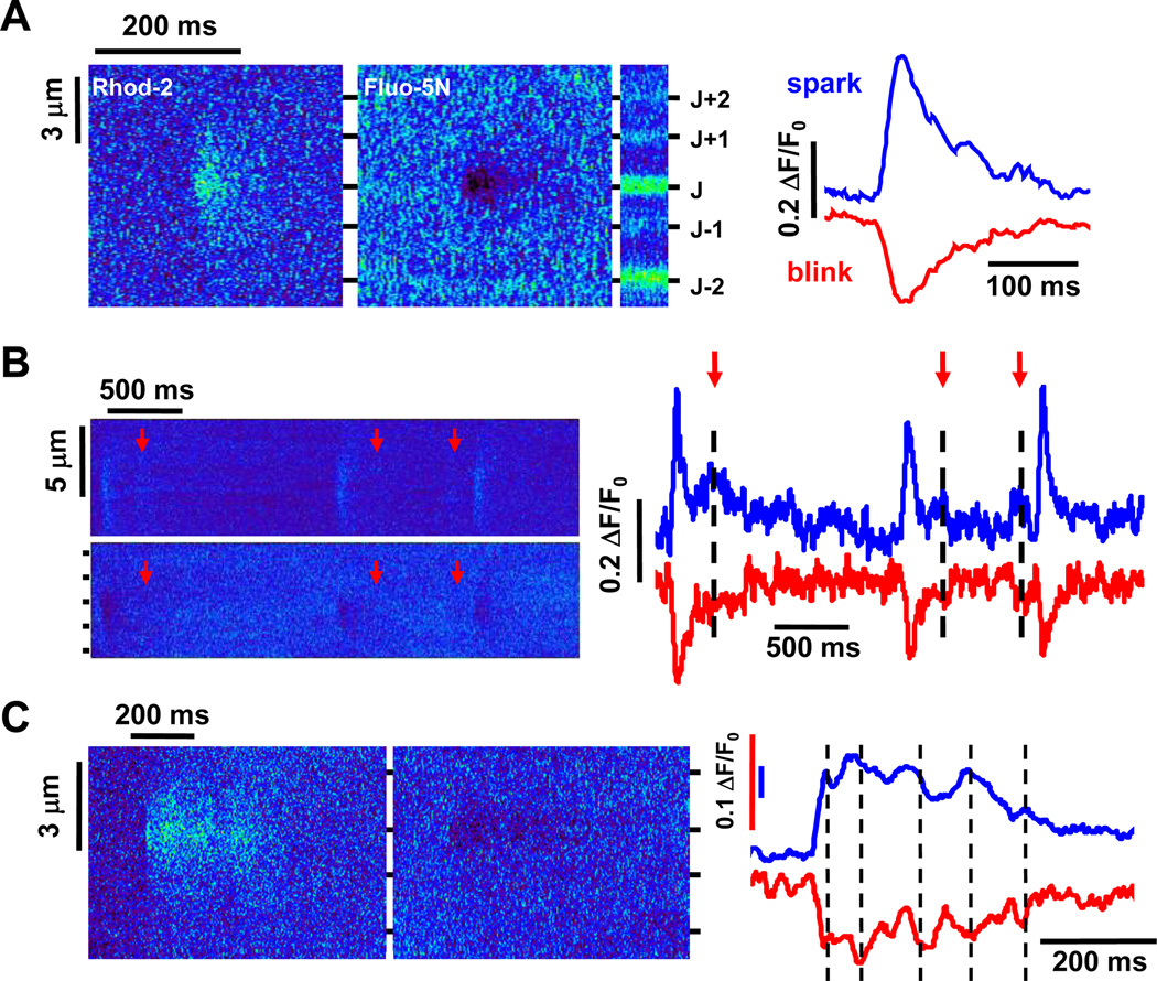
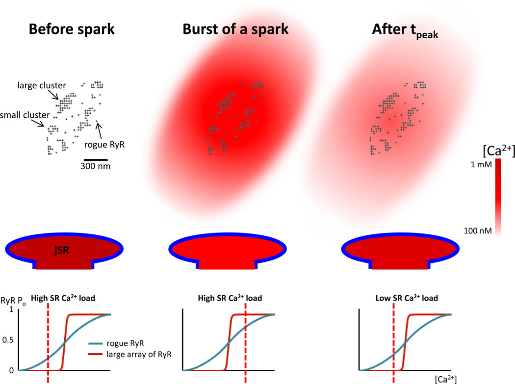
Ca(2+) release events underlie global Ca(2+) signaling yet they are regulated by local, subcellular signaling features. Here we review the latest developments of different elementary Ca(2+) release features that include Ca(2+) sparks, Ca(2+) blinks (the corresponding depletion of Ca(2+) in the sarcoplasmic reticulum (SR) during a spark) and the recently identified small Ca(2+) release events called quarky SR Ca(2+) release (QCR). QCR events arise from the opening of only a few type 2 ryanodine receptors (RyR2s) - possibly only one. Recent reports suggest that QCR events can be commingled with Ca(2+) sparks and may thus explain some variations observed in Ca(2+) sparks. The Ca(2+) spark termination mechanism and the number of RyR2 channels activated during a Ca(2+) spark will be discussed with respect to both Ca(2+) sparks and QCR events.


Figures

Fig 1
Fig 1. Examples of different elementary Ca2+ release events
A. Spark-blink pair. Linescan images of a Ca2+ spark (left panel) and its companion Ca2+ blink (mid left panel) after background subtraction with the corresponding time courses (right)in an intact rabbit ventricular myocyte. The enrichment of the fluo-5N dye in the junctional sarcoplasmic reticulum (jSR)can be seen on the unsubtracted fluo-5N image (mid-right). The spark-blink pair is centered on the jSR band identified as “J”. B. Quarky SR Ca2+ release (QCR) events. Succession of Ca2+ sparks and QCR events on the same jSR (top left) and their companion Ca2+ blinks and quarky SR Ca2+ depletion (QCD) events (bottom left),with the corresponding time courses (right). The arrows indicate the position of QCR and QCD events on the images and time course plots. C. Long spark-blink pair. An example of a long spark (left) and long blink (middle), with the corresponding time courses (right). The dashed lines on the traces show the correspondence between bumps on the spark and dips on the blink.
Fig 2
Fig 2. Chronology of the events happening during the time course of a Ca2+ spark
Organization of the different clusters of type 2 ryanodine receptors (RyR2s) at a jSR (top). Before the burst of a Ca2+ spark, all the RyR2s are in a closed state (top left column). The jSR is fully loaded of Ca2+ (mid left column) and the open probability of RyR2s is very low for large array and below 0.5 for rogue RyR2s (bottom left column). During the burst of a Ca2+ spark, the increase in cytosolic Ca2+ activates the large array of RyR2s (open probability very high) and most of the small arrays or rogue RyR2s (open probability superior at 0.5, bottom middle column). The [Ca2+] at the jSR reaches its nadir (mid middle column) whereas the [Ca2+] in the cytosol attains its peak (top middle column). Once the Ca2+ blink has reached its nadir, the low SR [Ca2+] has shifted the activation curves of the large array and rogue RyR2s to the right. The open probability of RyR2s is then very low for large array and below 0.5 for rogue RyR2s (bottom right column). Therefore, only some small arrays or rogue RyR2s will be activated during the tail of a spark (the large array also become refractory). The [Ca2+] in the SR will then increase (mid right column) at the same time as [Ca2+] in the cytosol will diminish (top right column).

Similar articles

Cited by

References

    1. Andrienko TN, Picht E, Bers DM. Mitochondrial free calcium regulation during sarcoplasmic reticulum calcium release in rat cardiac myocytes. J. Mol. Cell Cardiol. 2009;46:1027–1036. - PMC - PubMed
    1. Baddeley D, Jayasinghe ID, Lam L, Rossberger S, Cannell MB, Soeller C. Optical single-channel resolution imaging of the ryanodine receptor distribution in rat cardiac myocytes. Proc. Natl. Acad. Sci. U. S. A. 2009;106:22275–22280. - PMC - PubMed
    1. Bassani JW, Bassani RA, Bers DM. Relaxation in rabbit and rat cardiac cells: species-dependent differences in cellular mechanisms. J. Physiol. 1994;476:279–293. - PMC - PubMed
    1. Brochet DX, Xie W, Yang D, Cheng H, Lederer WJ. Quarky calcium release in the heart. Circ. Res. 2011;108:210–218. - PMC - PubMed
    1. Brochet DX, Yang D, Di Maio A, Lederer WJ, Franzini-Armstrong C, Cheng H. Ca2+ blinks: rapid nanoscopic store calcium signaling. Proc. Natl. Acad. Sci. U. S. A. 2005;102:3099–3104. - PMC - PubMed

Publication types

MeSH terms

Substances