

Bacteriophage t4 nanoparticles as materials in sensor applications: variables that influence their organization and assembly on surfaces
- PMID: 22454586
- PMCID: PMC3312445
- DOI: 10.3390/s90806298
Bacteriophage t4 nanoparticles as materials in sensor applications: variables that influence their organization and assembly on surfaces
Abstract
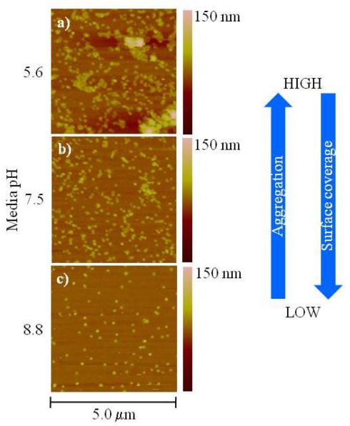
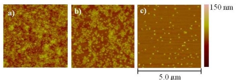
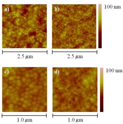
Bacteriophage T4 nanoparticles possess characteristics that make them ideal candidates as materials for sensors, particularly as sensor probes. Their surface can be modified, either through genetic engineering or direct chemical conjugation to display functional moieties such as antibodies or other proteins to recognize a specific target. However, in order for T4 nanoparticles to be utilized as a sensor probe, it is necessary to understand and control the variables that determine their assembly and organization on a surface. The aim of this work is to discuss some of variables that we have identified as influencing the behavior of T4 nanoparticles on surfaces. The effect of pH, ionic strength, substrate characteristics, nanoparticle concentration and charge was addressed qualitatively using atomic force microscopy (AFM).
Keywords: atomic force microscopy; bacteriophage T4; sensors.
Figures


References
-
- Fischlechner M., Donath E. Viruses as building blocks for materials and devices. Angew. Chem. Int. Ed. 2007;46:3184–3193. - PubMed
-
- Singh P., Gonzalez M.J., Manchester M. Viruses and their use in nanotechnology. Drug Dev. Res. 2006;67:23–41.
-
- Rao V.B. Methods and compositions comprising bacteriophage nanoparticles. Oct 13, 2005. U.S. Pat. Appl. Publ. Appl. No.: 11/015,294,
-
- Mattson T.L. Inviable virus particles as scaffolds for constructing flexible detection systems. Dec 31, 2002. U.S. Pat. 6,500,611,
-
- Szuchmacher Blum A., Soto C.M., Wilson C.D., Brower T.L., Pollack S.K., Schull T.L., Chatterji A., Lin T., Johnson J.E., Amsinck C., Franzon P., Shashidhar R., Ratna B.R. An engineered virus as a scaffold for three-dimensional self-assembly on the nanoscale. Small. 2005;1:702–706. - PubMed
LinkOut - more resources
Full Text Sources
Other Literature Sources
Miscellaneous
