. 2012 Mar 20:6:12.
doi: 10.3389/fncir.2012.00012. eCollection 2012.

Selective serotonergic excitation of callosal projection neurons

Affiliations

Selective serotonergic excitation of callosal projection neurons

Daniel Avesar et al. Front Neural Circuits. .

Abstract

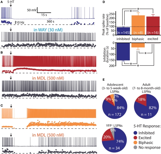
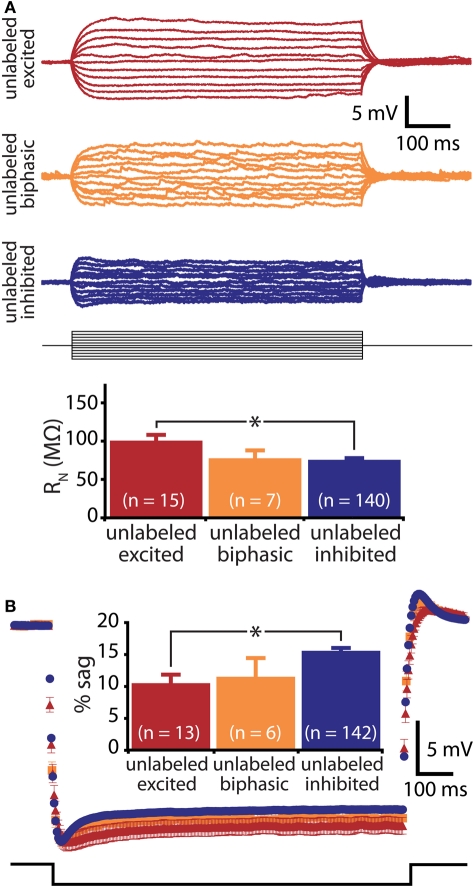
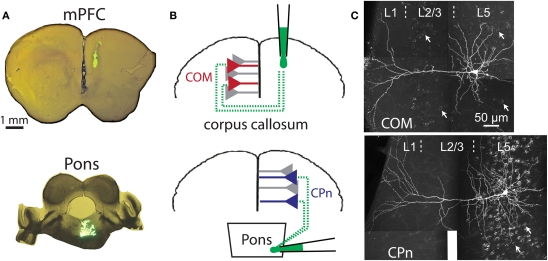
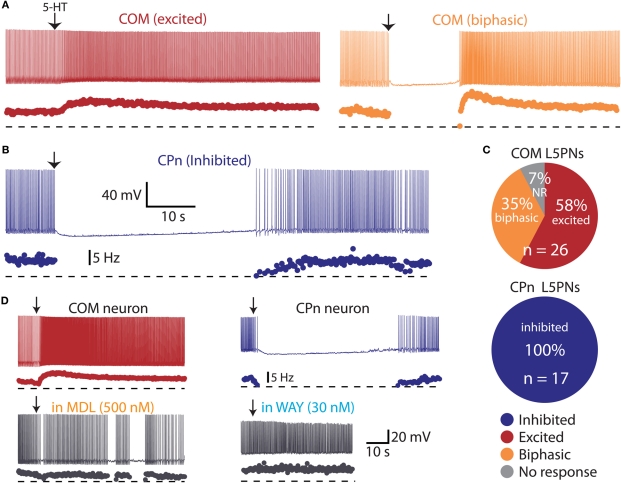
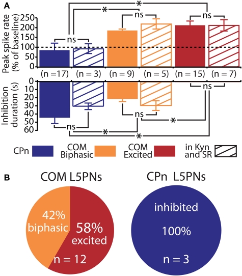
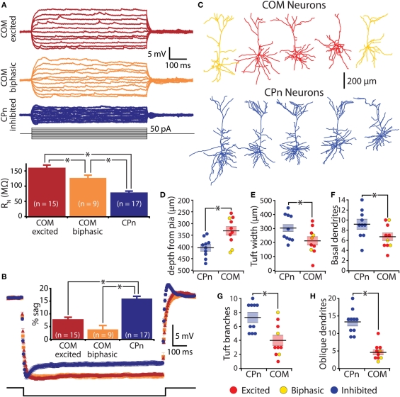
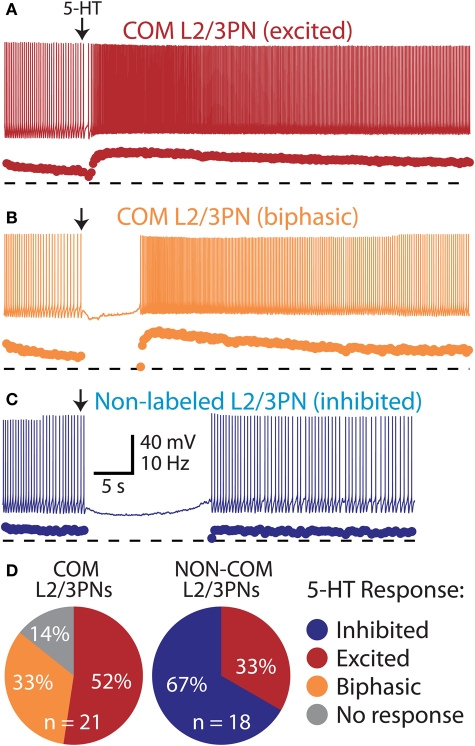
Serotonin (5-HT) acting as a neurotransmitter in the cerebral cortex is critical for cognitive function, yet how 5-HT regulates information processing in cortical circuits is not well understood. We tested the serotonergic responsiveness of layer 5 pyramidal neurons (L5PNs) in the mouse medial prefrontal cortex (mPFC), and found three distinct response types: long-lasting 5-HT(1A) (1A) receptor-dependent inhibitory responses (84% of L5PNs), 5-HT(2A) (2A) receptor-dependent excitatory responses (9%), and biphasic responses in which 2A-dependent excitation followed brief inhibition (5%). Relative to 5-HT-inhibited neurons, those excited by 5-HT had physiological properties characteristic of callosal/commissural (COM) neurons that project to the contralateral cortex. We tested whether serotonergic responses in cortical pyramidal neurons are correlated with their axonal projection pattern using retrograde fluorescent labeling of COM and corticopontine-projecting (CPn) neurons. 5-HT generated excitatory or biphasic responses in all 5-HT-responsive layer 5 COM neurons. Conversely, CPn neurons were universally inhibited by 5-HT. Serotonergic excitation of COM neurons was blocked by the 2A antagonist MDL 11939, while serotonergic inhibition of CPn neurons was blocked by the 1A antagonist WAY 100635, confirming a role for these two receptor subtypes in regulating pyramidal neuron activity. Selective serotonergic excitation of COM neurons was not layer-specific, as COM neurons in layer 2/3 were also selectively excited by 5-HT relative to their non-labeled pyramidal neuron neighbors. Because neocortical 2A receptors are implicated in the etiology and pathophysiology of schizophrenia, we propose that COM neurons may represent a novel cellular target for intervention in psychiatric disease.

Keywords: 5-HT1A receptor; 5-HT2A receptor; cerebral cortex; corpus callosum; neocortex; pyramidal neuron; retrograde labeling; serotonin.


Figures

Figure 1
Figure 1
Diversity of serotonin responses in prefrontal layer 5 pyramidal neurons (L5PNs). (A–C) Responses to focal serotonin application (100 μM, 10 s, beginning at arrow) in L5PNs during periods of current-driven action potential generation. Three response types were observed: inhibition, blocked by the 1A antagonist WAY 100635 (A), excitation, blocked by the 2A antagonist MDL 11939 (B), and biphasic responses consisting of short-duration inhibition followed by excitation, also sensitive to the 2A antagonist (C). (D) Comparisons of the duration of inhibition (bottom) and peak changes in spike rate (top) according to serotonergic response type. (E) Proportions of L5PNs exhibiting different serotonin responses in neurons from the mPFCs of adolescent mice, fully adult mice, and adolescent Thy1-YFP mice in which a subpopulation of L5PNs are selectively labeled with yellow fluorescent protein (YFP).
Figure 2
Figure 2
Responsiveness to serotonin correlates with membrane physiology. (A) Top, voltage responses to somatic current injections (−50 to 50 pA in 5 pA steps) in unlabeled L5PNs. Bottom, comparison of input resistance (RN) in neurons having different responses to serotonin. (B) Average (± SEM) voltage responses to hyperpolarizing current injections sufficient to generate a peak hyperpolarization of ∼20 mV in unlabeled L5PNs grouped according to their responsiveness to serotonin (blue indicates serotonergic inhibition, red indicates serotonergic excitation, and orange indicates biphasic responses). Comparison of results is inset. Asterisks indicate p < 0.05.
Figure 3
Figure 3
Retrograde labeling of prefrontal projection neurons. (A) Images of coronal sections of prefrontal cortex (top) or brainstem (bottom) showing locations of dye injection for retrograde labeling of callosal/commisseral (COM) and corticopontine (CPn) projection neurons. (B) Cartoons of retrograde labeling of COM (top) and CPn neurons (bottom). (C) Images of COM (top) and CPn (bottom) neurons that were filled with biocytin during recording and labeled with a fluorescent-conjugated avidin. The punctate Retrobead staining of other COM and CPn somata are visible in the backgrounds (white arrows point out examples).
Figure 4
Figure 4
Serotonin selectively excites commissural/callosal (COM) projection neurons. (A) Excitatory (left) and biphasic (right) responses to focal serotonin application in COM neurons. (B) Inhibitory response to serotonin in a corticopontine (CPn) neuron. (C) Proportion of COM and CPn neurons excited (red), inhibited (blue), or having biphasic (orange) responses to 5-HT. Non-responsive neurons (NR) shown in gray. (D) Serotonergic excitation of COM neurons (left) and inhibition of CPn neurons (right) was blocked by antagonists specific for 2A (MDL 11939) or 1A (WAY 100635) receptors, respectively.
Figure 5
Figure 5
Serotonergic regulation of commissural/callosal (COM) and corticopontine (CPn) projection neurons does not require changes in fast synaptic transmission. (A) Comparisons of the duration of inhibition (bottom) and serotonin-induced changes in spike rate (top) in COM and CPn neurons in regular saline (solid bars) and in the presence of synaptic blockers (4 mM kynurenic acid, 10 μM SR 95531; striped bars). NS indicates non-significant differences between within-cell-type groups (unpaired Student's t-tests). Asterisks indicate p < 0.05 (One-Way ANOVAs). (B) Proportions of serotonergic responses in COM (top) and CPn neurons (bottom) recorded in the presence of 4 mM kynurenic acid and 10 μM SR-95531.
Figure 6
Figure 6
Physiological and morphological differences in commissural/callosal (COM) and corticopontine (CPn) projection neurons. (A) Top, responses to somatic current injections in retrograde-labeled COM and CPn neurons. Bottom, comparison of input resistance (RN) in neurons having different responsiveness to 5-HT. (B) Average (± SEM) voltage responses to hyperpolarizing current injections sufficient to generate a peak hyperpolarization of ∼20 mV in labeled COM and CPn neurons grouped according to serotonin response (blue indicates inhibition, red indicates excitation, and orange indicates biphasic responses). Comparison of results is inset. (C) Representative morphologies of COM and CPn neurons. (D–H) Comparisons of morphological properties in COM and CPn neurons (n = 10 for each group). Response type of the neuron is color-coded, as indicated. Asterisks indicate p < 0.05.
Figure 7
Figure 7
Selective excitation of pyramidal neurons in layers 2/3 of the mPFC.(A) Excitatory response to 5-HT in a COM-labeled L2/3PN (voltage trace, above, and instantaneous spike frequency [ISF] plot, below). Dashed lines indicate zero Hz. (B) Serotonergic response in a “biphasic” COM L2/3PN. (C) Serotonergic inhibition of a non-labeled L2/3PN in tissue labeled for COM neurons. (D) Proportions of serotonergic responses in COM-labeled and neighboring non-labeled L2/3PNs.

References

    1. Aghajanian G. K., Marek G. J. (1997). Serotonin induces excitatory postsynaptic potentials in apical dendrites of neocortical pyramidal cells. Neuropharmacology 36, 589–599 10.1074/jbc.270.40.23823 - DOI - PubMed
    1. Amargos-Bosch M., Bortolozzi A., Puig M. V., Serrats J., Adell A., Celada P., Toth M., Mengod G., Artigas F. (2004). Co-expression and in vivo interaction of serotonin1A and serotonin2A receptors in pyramidal neurons of prefrontal cortex. Cereb. Cortex 14, 281–299 10.1093/cercor/bhg128 - DOI - PubMed
    1. Araneda R., Andrade R. (1991). 5-Hydroxytryptamine2 and 5-hydroxytryptamine 1A receptors mediate opposing responses on membrane excitability in rat association cortex. Neuroscience 40, 399–412 10.1016/0306-4522(91)90128-B - DOI - PubMed
    1. Arlotta P., Molyneaux B. J., Chen J., Inoue J., Kominami R., Macklis J. D. (2005). Neuronal subtype-specific genes that control corticospinal motor neuron development in vivo. Neuron 45, 207–221 10.1016/j.neuron.2004.12.036 - DOI - PubMed
    1. Beique J. C., Campbell B., Perring P., Hamblin M. W., Walker P., Mladenovic L., Andrade R. (2004). Serotonergic regulation of membrane potential in developing rat prefrontal cortex: coordinated expression of 5-hydroxytryptamine (5-HT)1A, 5-HT2A, and 5-HT7 receptors. J. Neurosci. 24, 4807–4817 10.1523/JNEUROSCI.5113-03.2004 - DOI - PMC - PubMed

LinkOut - more resources