

Non-Hodgkin's Lymphoma Presenting as a Huge Ocular Adnexal and Forehead Mass
- PMID: 22454706
- PMCID: PMC3306071
Non-Hodgkin's Lymphoma Presenting as a Huge Ocular Adnexal and Forehead Mass
Abstract
Purpose: To report a case of non-Hodgkin's lymphoma (NHL) presenting as an ocular adnexal and forehead mass.
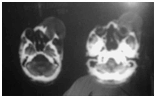
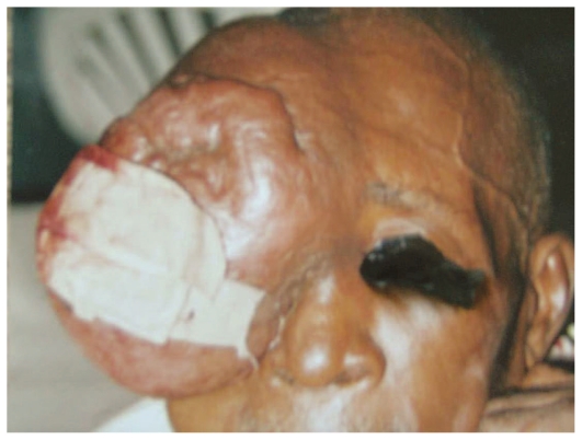
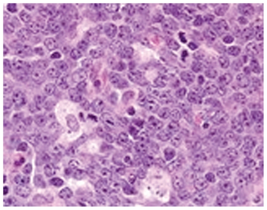
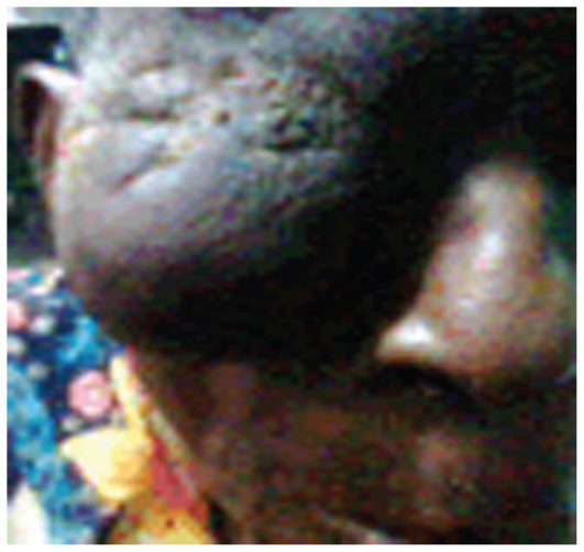
Case report: An elderly male patient was referred by a neurosurgeon to the eye clinic with a six-month history of a massive tumor measuring 12×16×8 cm involving the right side of the forehead, eyebrow and upper eyelid. Neurological examination had been normal and computed tomography revealed no intracranial extension. The patient was referred to an otorhinolaryngologist who performed an incisional biopsy which revealed the mass to be NHL. He received chemotherapy with CHOP regimen (cyclophosphamide, adriamycin, vincristine and prednisolone) resulting in reduction in lesion size leaving a phthysical eyeball and a ptotic lid.
Conclusion: Non-Hodgkin's lymphoma may occur in almost any part of the body and should be considered in the differential diagnosis of extralymphoid tumors.
Keywords: Forehead Mass; Lid Mass; Non-Hodgkin’s Lymphoma.
Figures
References
-
- Hernández-Rivera G, Aguayo-González A, Cano-Castellanos R, Loarca-Piña LM. Current therapeutic advances in the treatment of non-Hodgkin lymphoma. Gac Med Mex. 2008;144:275–277. - PubMed
-
- Omoti AE, Omoti CE. Ophthalmic manifestations of lymphoma. Ann Afri Med. 2007;6:89–93. - PubMed
-
- Coupland SE, Foss HD, Assaf C, Auw-Haedrich C, Anastassiou G, Anagnostopoulos I, et al. T-cell and T/natural killer-cell lymphomas involving ocular and ocular adnexial tissues: a clinicopathologic, immunohistochemical, and molecular study of seven cases. Ophthalmology. 1999;106:2109–2120. - PubMed
-
- Schmidt-Erfurth U, Bastian GO, Bopp S, Lucke K, Laqua H. Clinical heterogeneity of non-Hodgkin’s lymphoma of the eye with extraocular manifestations. Ophthalmologe. 1994;91:357–363. - PubMed
Publication types
LinkOut - more resources
Full Text Sources
Research Materials