. 2011 Oct;6(4):241-8.

Indocyanine green angiography guided management of vogt-koyanagi-harada disease

Affiliations

Indocyanine green angiography guided management of vogt-koyanagi-harada disease

Nadia Bouchenaki et al. J Ophthalmic Vis Res. 2011 Oct.

Abstract

Purpose: To report the management of Vogt-Koyanagi-Harada (VKH) disease based on indocyanine green angiography (ICGA).

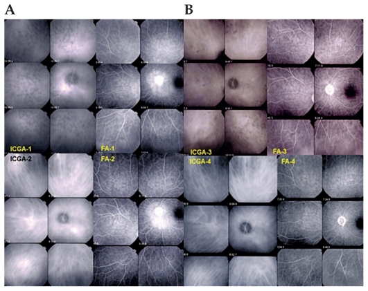
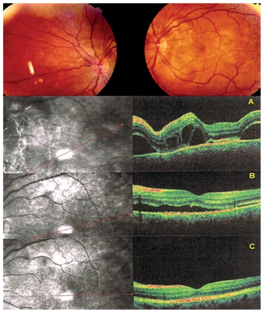
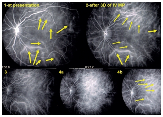
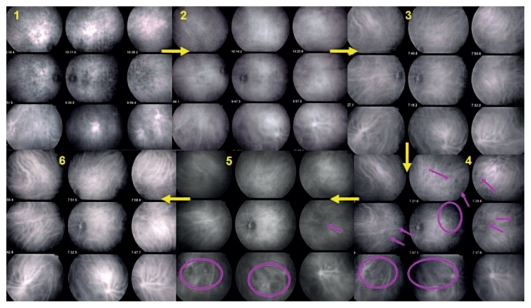
Methods: VKH patients with acute episodes of inflammation (inaugural or recurrent) who had received standard ICGA-guided care were studied retrospectively. Standard of care included high dose systemic corticosteroids at presentation and close ICGA follow-up with addition of immunosuppressive agents and/or intensification of ongoing therapy when recurrent choroidal lesions were detected by ICGA. Visual acuity, number of subclinical recurrences, type and duration of therapy, proportion of quiescent patients after therapy, and ICGA findings were recorded.

Results: Nine patients including 8 female and one male subject were studied. Five patients had inaugural disease and 4 presented with recurrent acute episodes. Visual acuity increased from 0.86±0.36 to 1.14±0.34 in the right eyes, and from 0.77±0.34 to 1.05±0.33 in the left eyes. The number of ICGA-detected occult choroidal recurrences amounted to 13. Mean duration of treatment was 30.1±34.6 months leading to recurrence-free status after discontinuation of therapy in 6 cases with mean duration of 29.5 months.

Conclusion: Continuous monitoring and aggressive therapy guided by ICGA in VKH disease prolongs treatment as compared to textbook guidelines but offers the prospect of reaching inflammation-free status after discontinuation of therapy. Zero tolerance to subclinical choroidal inflammation avoids irremediable evolution towards sunset glow fundus in patients treated early after the initial acute inflammatory attack.

Keywords: Indocyanine Green Angiography; Therapy; Vogt-Koyanagi-Harada Disease.


Figures

Figure 1
Figure 1
(A) An example of ICGA-guided management of VKH. A patient with an inaugural episode of VKH disease shows numerous HDDs and complete vessel fuzziness (top left frames, ICGA-1). FA (top right frames, FA-1) shows disc hyperfluorescence and peripapillary exudative retinal detachment. After intravenous methylprednisolone and oral prednisone, there is complete disappearance of HDDs and fuzziness of choroidal vessels, the pattern of which is clearly seen again (bottom left frames, ICGA-2), while there is residual disc and peripapillary hyperfluorescence (bottom right frames, FA-2). (B) After prednisone tapering, subclinical recurrence of choroidal inflammation with reappearance of HDDs and fuzziness of choroidal vessels (top left frames, ICGA-3) are seen, while FA is quasi normal with only residual disc hyperfluorescence (top right frames, FA-3). After increase of oral prednisone and addition of mycophenolate mofetil, the choroidal inflammatory lesions (HDDs) disappeared two months later and choroidal vessels regained a normal appearance (bottom left frames, ICGA-4). ICGA, indocyanine green angiography VKH, Vogt-Koyanagi-Harada HDDs, hypofluorescent dark dots FA, fluorescein angiography
Figure 2
Figure 2
Fundus and OCT findings in acute VKH. Fundus signs showed bilateral serous retinal detachments in this patient (only the left eye is shown) diagnosed 7 days after the onset of symptoms. OCT illustrates subretinal fluid at presentation (top scan, A). After 3 days of intravenous methylprednisolone, the amount of fluid has decreased substantially (middle scan, B) with quasi normalization after one month of treatment (bottom scan, C). OCT, optical coherence tomography VKH, Vogt-Koyanagi-Harada
Figure 3
Figure 3
Early choroidal hyperfluorescent vessels three days after administration of intravenous methylprednisolone (top right) in a patient with an acute inaugural VKH episode at presentation (top left). Normalization of choroidal vessels after 3 months of combined prednisone and mycophenolate mofetil therapy (bottom left, 3). Reappearance of early hyperfluorescent choroidal vessels, together with HDDs 6 weeks later upon discontinuation of prednisone (bottom frames 4a and 4b). VKH, Vogt-Koyanagi-Harada HDDs, hypofluorescent dark dots
Figure 4
Figure 4
Numerous HDDs and fuzzy vessels at presentation (1). Decrease in HDDs after one month of prednisone therapy with persistence of fuzzy vessels observed with ICGA-guided follow-up (2). Choroidal inflammation finally responded to combined prednisone and mycophenolate mofetil therapy resulting in resolution of HDDs and restoration of normal pattern of choroidal vessels (3). Six weeks later and after discontinuation of prednisone, recurrence of occult choroidal inflammation was noted with reappearance of HDDs and vessel fuzziness in some areas of the fundus (4, crimson circles and arrows) which slowly responded to the addition of cyclosporine after two months (5) leading to a quasi normal choroidal appearance after another two months (6). ICGA, indocyanine green angiography HDDs, hypofluorescent dark dots

References

    1. Bouchenaki N, Herbort CP. Stromal choroiditis. In: Pleyer U, Mondino B, editors. Essentials in Ophthalmology: Uveitis and Immunological Disorders. 1st ed. Berlin, Heidelberg, New York: Springer; 2004. pp. 234–253.
    1. Sugita S, Takase H, Taguchi C, Imai Y, Kamoi K, Kawaguchi T, et al. Ocular infiltrating CD4+ T cells from patients with Vogt-Koyanagi-Harada disease recognize human melanocyte antigens. Invest Ophthalmol Vis Sci. 2006;47:2547–2554. - PubMed
    1. Damico FM, Cunha-Neto E, Goldberg AC, Iwai LK, Marin ML, Hammer J, et al. T-cell recognition and cytokine profile induced by melanocyte epitopes in patients with HLA-DRB1*0405-positive and -negative Vogt-Koyanagi-Harada uveitis. Invest Ophthalmol Vis Sci. 2005;46:2465–2471. - PubMed
    1. Sonoda S, Nakao K, Ohba N. Extensive chorioretinal atrophy in Vogt-Koyanagi-Harada disease. Jpn J Ophthalmol. 1999;43:113–119. - PubMed
    1. Rao NA. Treatment of Vogt-Koyanagi-Harada disease by corticosteroids and immunosuppressive agents. Ocul Immunol Inflamm. 2006;14:71–72. - PubMed

LinkOut - more resources