. 2011 Oct;6(4):270-83.

Myopia and inflammation

Affiliations

Myopia and inflammation

Carl P Herbort et al. J Ophthalmic Vis Res. 2011 Oct.

Abstract

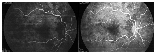
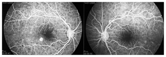
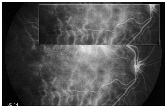
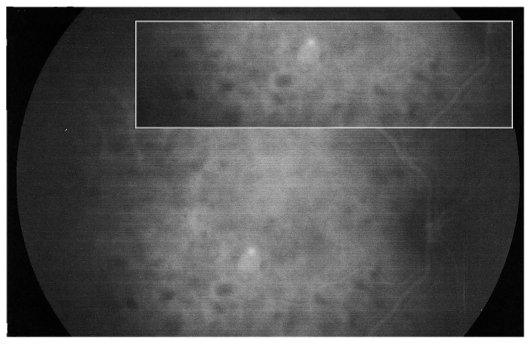
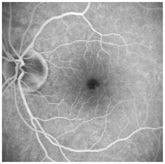
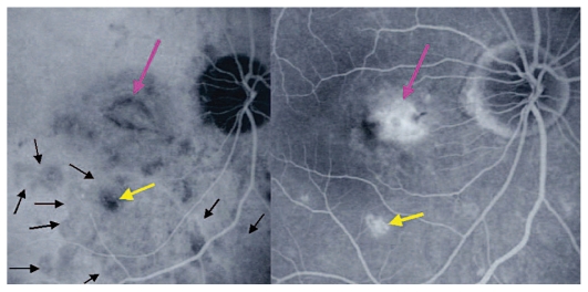
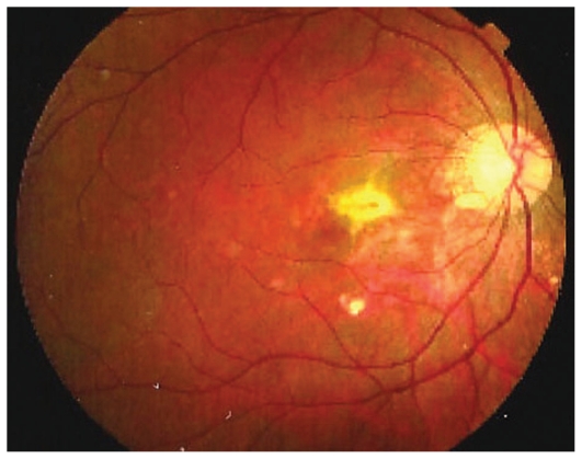
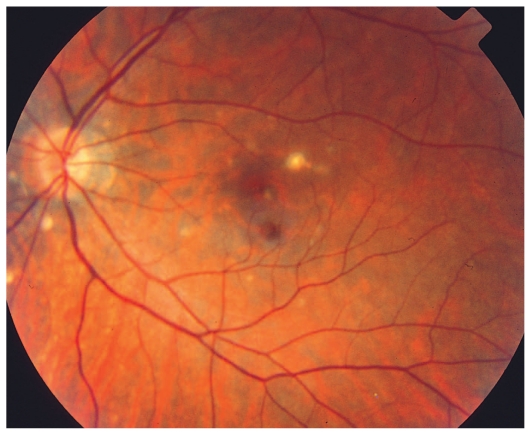
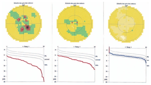
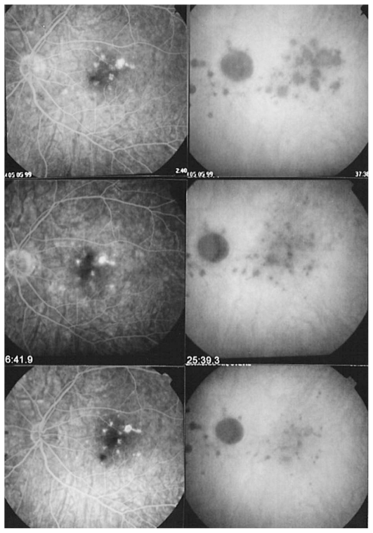
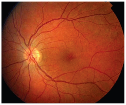
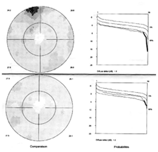
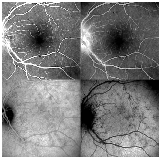
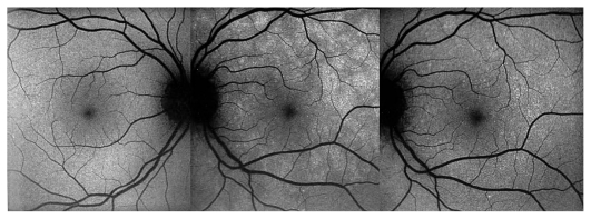
The correlation between myopia and intraocular inflammation has rarely been explored. The aim of this article is to review myopic changes induced by inflammatory diseases and inflammatory diseases related to myopia, followed by a discussion on inflammatory choroidal neovascularization. Clinical cases are used to illustrate these conditions. The review does not include inflammatory conditions caused by surgical interventions employed for treatment of myopia. Uveitic conditions that can induce a myopic shift include sclero-choroidal inflammation, lens induced myopia due to steroid cataracts, juvenile idiopathic arthritis (JIA) induced myopia, and transient drug induced myopia due to sulfonamides and acetazolamide used for treatment of ocular toxoplasmosis and inflammatory cystoid macular edema, respectively. Most inflammatory conditions related to myopia are conditions involving the choriocapillaris. These include multifocal choroiditis and/or punctate inner choroiditis, multiple evanescent white dot syndrome and acute idiopathic blind spot enlargement. It can be hypothesized that fragility of the choriocapillaris due to particular anatomic changes due to myopia, together with unknown immunogenetic factors predispose myopic eyes to primary inflammatory choriocapillaropathies.

Keywords: Choriocapillaritis; Inflammation; Multifocal Choroiditis; Multiple Evanescent White Dot Syndrome; Myopia; Vogt-Koyanagi-Harada Disease.


Figures

Figure 1A
Figure 1A
Hyperacute VKH, nonhomogeneous filling of the choriocapillaris.
Figure 1B
Figure 1B
Hyperacute VKH, patchy hyperfluorescence on FA.
Figure 1C
Figure 1C
Hyperacute VKH disease, early hyperfluorescence of choroidal vessels and leakage from choroidal stromal vessels indicating choroidal vasculitis.
Figure 1D
Figure 1D
Hyperacute VKH disease, note numerous regularly sized and evenly distributed hypofluorescent dark dots on a background of diffuse choroidal hyperfluorescence, indicating granulomas of the choroid.
Figure 1E
Figure 1E
Hyperacute VKH disease, exudative retinal detachments shown on ICGA in the left eye. Note the numerous dark dots indicating choroidal granulomas.
Figure 1F
Figure 1F
Hyperacute VKH disease, UBM image shows supraciliary effusion explaining myopization.
Figure 2A
Figure 2A
Fundus appearance in multifocal choroiditis (MFC). Right and left fundus frames of a case of MFC showing typical dispersed lesions. No sign of disease recurrence appears in the left eye.
Figure 2B
Figure 2B
ICGA in multifocal choroiditis. ICGA shows widespread non-perfusion in the left eye not apparent on fundus photographs (Fig. 2A) nor on FA (Fig. 2C). Only ICGA allows detection of recurrence, an example of ICGA “iceberg” and “submarine” concepts indicating that ICGA signs can be pronounced despite minimal or absent fundus or FA signs. Note also the extensive ischemia which explains the high percentage of choroidal neovascularization in MFC. Right eye is currently inactive. (Courtesy of Alessandro Mantovani, Como, Italy).
Figure 2C
Figure 2C
FA in multifocal choroiditis. As shown in figure 2B, extensive ICGA signs can be present in the absence of fundus or FA signs.
Figure 2D
Figure 2D
Choroidal neovascularization complicating MFC shown on ICGA (left) and FA (right). As shown in figure 2B, choriocapillaris ischemia is usually extensive in MFC and can only be detected by ICGA. This figure shows the development of CNV in the right eye (crimson arrow) concomitant with an acute recurrence of MFC causing extensive choriocapillaris ischemia (black arrows) not visible on FA. The darker areas are old cicatricial foci that can also be seen on FA (window defects, yellow arrows). (Courtesy of Alessandro Mantovani, Como, Italy)
Figure 2E
Figure 2E
Cicatricial CNV treated by photodynamic therapy in MFC. The old cicatricial lesions that were seen on ICGA (very dark spots) and FA (window defect hyperfluorescence) are well identified.
Figure 3A
Figure 3A
Punctate inner choroiditis, fundus image of the left eye shows one larger chorioretinal scar and several faint choroiditis foci. New recurrent lesions are not detectable.
Figure 3B
Figure 3B
Octopus visual field at presentation and during follow-up. Extensive centrally coalescent scotomata at presentation (left). Significant improvement after one month of corticosteroid therapy (middle) and complete recovery after six months of treatment (right). (Source of illustration: reference 18)
Figure 3C
Figure 3C
Evolution of FA (left column) and ICGA lesions (right column) at presentation and under corticosteroid treatment for punctate inner choroidopathy. At presentation (top images), FA only shows cicatricial lesions from old foci (window defects) and does not reveal the extensive occult recurrence shown by ICGA. After one month of systemic therapy, FA frames show the same cicatricial lesions (middle left) whereas the fresh lesions have regressed on ICGA (middle right). After 6 months of corticosteroid therapy (bottom images), FA shows the same appearance of old cicatricial lesions whereas ICGA shows complete regression of the fresh lesions; old atrophic scars due to episodes are also visible on FA frames.
Figure 4A
Figure 4A
Fundus image of the left eye in a patient with MEWDS. The fundus is almost normal with only a slightly nonhomogeneous appearance in the fovea.
Figure 4B
Figure 4B
Octopus visual fields in MEWDS. Slight increase in mean defect (MD) and shallow relative scotoma at presentation (top image) with normalization 7 weeks later during the healing phase.
Figure 4C
Figure 4C
FA and ICGA frames in a patient with MEWDS. FA (top images) shows tiny hyperfluorescent spots in the posterior pole and disc leakage on the late frame (top right). ICGA (bottom images) shows extensive areas of choriocapillaris non- perfusion in the posterior pole, well visible in the intermediate phase (bottom left) but much clearly outlined in the late phase (bottom right).
Figure 4D
Figure 4D
Fundus auto-fluorescence (FAF) in MEWDS. Geographic and confluent areas of increased FAF in the left diseased eye (middle picture) corresponding to dark ICGA areas which almost completely recovered in the healing stage (right image).

References

    1. Nomura H, Ando F, Nino N, Shimokata H, Miyake Y. The relationship between intraocular pressure and refractive error adjusting for age and central corneal thickness. Ophthalmic Physiol Opt. 2004;24:41–45. - PubMed
    1. Xu L, Wang Y, Wang S, Wang Y, Jonas JB. High myopia and glaucoma susceptibility: the Beijing eye study. Ophthalmology. 2007;114:216–220. - PubMed
    1. Lee YA, Shih YF, Lin LL, Huang JY, Wang TH. Association between high myopia and progression of visual field loss in primary open-angle glaucoma. J Formos Med Assoc. 2008;107:952–957. - PubMed
    1. Bremer F. Origin of corticosteroid glaucoma. Bull Soc Belge Ophtalmol. 2007;304:111–116. - PubMed
    1. Gross SA, von Noorden GK, Jones DB. Necrotizing scleritis and transient myopia following strabismus surgery. Ophthalmic Surg. 1993;24:839–841. - PubMed

LinkOut - more resources