

Idiopathic retinal vasculitis, aneurysms and neuroretinitis syndrome associated with positive perinuclear antineutrophil cytoplasmic antibody
- PMID: 22454754
- PMCID: PMC3306109
Idiopathic retinal vasculitis, aneurysms and neuroretinitis syndrome associated with positive perinuclear antineutrophil cytoplasmic antibody
Abstract
Purpose: To report a case of idiopathic retinal vasculitis, aneurysms and neuroretinitis (IRVAN) syndrome associated with positive perinuclear antineutrophil cytoplasmic antibody (P-ANCA).
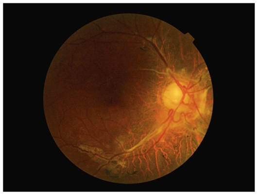
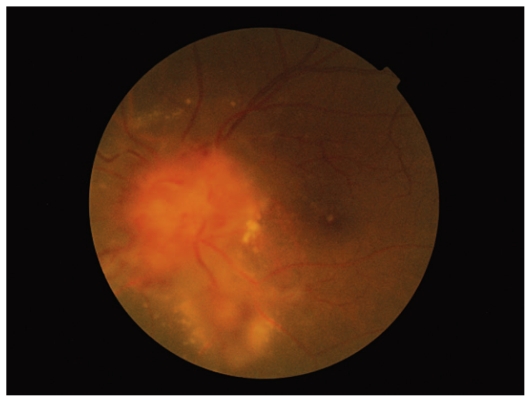
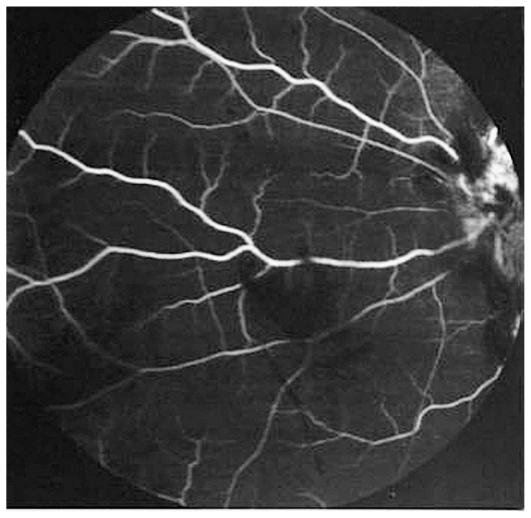
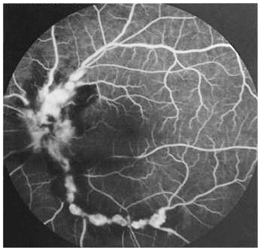
Case report: A 51-year-old man presented with loss of vision in his right eye since many years ago and blurred vision in his left eye over the past year. Ophthalmologic examination revealed optic atrophy and old vascular sheathing in the right eye and blurred disc margin, macular exudation, flame shaped hemorrhages, retinal vascular sheathing and multiple aneurysms at arterial bifurcation sites in the left eye, findings compatible with IRVAN syndrome. On systemic workup, the only notable finding was P-ANCA positivity.
Conclusion: IRVAN syndrome may be a retinal component of P-ANCA associated vasculitis.
Keywords: IRVAN Syndrome; P-ANCA.
Figures
References
-
- Chang TS, Aylward GW, Davis JL, Mieler WF, Oliver GL, Maberley AL, et al. Idiopathic retinal vasculitis, aneurysms and neuroretinitis. Ophthalmology. 1995;102:1089–1097. - PubMed
-
- Samuel MA, Equi RA, Chang TS, Mieler W, Jampol LM, Hay D, et al. Idiopathic retinitis, vasculitis, aneurysms, and neuroretinitis (IRVAN): new observations and a proposed staging system. Ophthalmology. 2007;114:1526–1529. - PubMed
-
- Soheilian M, Nourinia R, Tavallali A, Peyman GA. Idiopathic Retinal Vasculitis, Aneurysms, and Neuroretinitis Syndrome Associated with Positive Perinuclear Antineutrophil Cytoplasmic Antibody (P-ANCA) Retinal Cases & Brief Reports. 2010;4:198–201. - PubMed
-
- Mayet WJ, Helmreich-Becker I, Meyer zum Büschenfelde KH. The pathophysiology of anti-neutrophil cytoplasmic antibodies (ANCA) and their clinical relevance. Crit Rev Oncol Hematol. 1996;23:151–156. - PubMed
-
- Kincaid J, Schatz H. Bilateral retinal arteritis with multiple aneurysmal dilatations. Retina. 1983;3:171–178. - PubMed
Publication types
LinkOut - more resources
Full Text Sources
Molecular Biology Databases
Miscellaneous