Review
. 2012 Mar 28:10:24.
doi: 10.1186/1477-7827-10-24.

hCG, the wonder of today's science

Affiliations
Review

hCG, the wonder of today's science

Laurence A Cole. Reprod Biol Endocrinol. .

Abstract

Background: hCG is a wonder. Firstly, because hCG is such an extreme molecule. hCG is the most acidic glycoprotein containing the highest proportion of sugars. Secondly, hCG exists in 5 common forms. Finally, it has so many functions ranging from control of human pregnancy to human cancer. This review examines these molecules in detail.

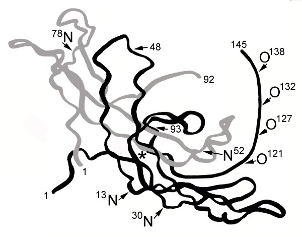
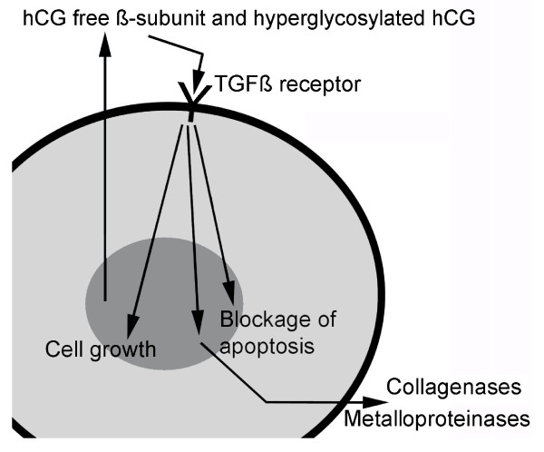
Content: These 5 molecules, hCG, sulfated hCG, hyperglycosylated hCG, hCG free beta and hyperglycosylated free beta are produced by placental syncytiotrophoblast cells and pituitary gonadotrope cells (group 1), and by placental cytotrophoblast cells and human malignancies (group 2). Group 1 molecules are both hormones that act on the hCG/LH receptor. These molecules are central to human menstrual cycle and human pregnancy. Group 2 molecules are autocrines, that act by antagonizing a TGF beta receptor. These molecules are critical to all advanced malignancies.

Conclusions: The hCG groups are molecules critical to both the molecules of pregnancy or human life, and to the advancement of cancer, or human death.


Figures

Figure 1
Figure 1
The three dimensional structure of deglycosylated hCG as shown by X-ray defraction [4]. O and N mark O-linked and N-linked oligosaccharides and * marks the site of the cystine knot, common to TGFß. Grey is α- and black is ß-subunit.
Figure 2
Figure 2
Formation of villous trophoblast. A. Cytotrophoblast columns in early implanted embryo. B. Extension of columns and differentiation of peripheral cells. B. and C. Folding of extensions caused by shape of syncytiotrophoblast cells. C and D formation of trophoblastic villi. No vascular supply, spiral arteries or fetal vasculature is shown.
Figure 3
Figure 3
Human placental hemochorial placentation. While hCG and hyperglycosylated hCG force villous trophoblast tissue formation [13-17], hCG promotes the development and growth of uterine spiral arteries [19-26]. Angiogenesis forces the protrusion of arteries to reach invading villous trophoblast tissue [19-26]. hCG also promotes the formation of the umbilical circulation in villous tissue and the formation of the umbilical cord [27-32]. All linked together, villous trophoblast tissue, maternal spiral artery blood and fetal umbilical circulation and you have hemochorial placentation, efficient fetal nutrient exchange, as illustrated. In hemochorial placentation, spiral artery bring maternal blood into one of 4-7 hemochorial placentation chambers. Blood fills the chamber, nutrients (oxygen/glucse/amino acids) them pass across syncytiotrophoblast cells into villous side-arms or floating villi. They are then rapidly absorbed by the umbilical circulation.
Figure 4
Figure 4
The three dimensional structure of hCG [4]. In order to form crystals hCG was first deglycosylated. hCG and hyperglycosylated hCG have identical amino acid sequence [1], yet vary in function. hCG binds the hCG/LH receptor while hyperglycosylated hCG antagonizes a TGFß receptor [6-8]. As such the glycosylation (all removed) has to make a structural difference to the two hCG forms. This difference is not shown. Oligosaccharide are added to the three dimensional structure assuming the carbohydrate structure observation of Elliott et al. [1]. Oligosaccharides are charged so must project towards the surface of the molecule. L in oligosaccharides is N-acetylgalactosamine, A is N-acetylglucosamine, S is sialic acid or N-acetyl-neuraminic acid, G is galactose, M is mannose and F is fucose.
Figure 5
Figure 5
The three dimensional structure of hyperglycosylated hCG [4]. In order to form crystals hCG was first deglycosylated. hCG and hyperglycosylated hCG have identical amino acid sequence [1], yet vary in function. hCG binds the hCG/LH receptor while hyperglycosylated hCG antagonizes a TGFß receptor [6-8]. As such the glycosylation (all removed) has to make a structural difference to the two hCG forms. This difference is not shown. Oligosaccharide are added to the three dimensional structure assuming the carbohydrate structure observation of Elliott et al. [1]. Oligosaccharides are charged so must project towards the surface of the molecule. L in oligosaccharides is N-acetylgalactosamine, A is N-acetylglucosamine, S is sialic acid or N-acetyl-neuraminic acid, G is galactose, M is mannose and F is fucose.
Figure 6
Figure 6
Proposed pathways of free ß-subunit and hyperglycosylated hCG in advanced cancer cases. As illustrated, free ß-subunit and hyperglycosylated hCG are hCG variants with exposed TGFß binding structures [1-4,6], these are autocrines which antagonize the TGFß receptor, promoting cell growth and blocking cell apoptosis. As a result of the antagonism, collagenases and metalloproteinases are produced by cells [94,95]. As illustrated, cells secrete hCG free ß-subunit or hyperglycosylated hCG. This enters the circulation and rotates around the body. hCG free ß-subunit of hyperglycosylated hCG then bind back on a receptor on the cancer cells, a TGFß receptor, and antagonize this receptor. As a result cell growth is promoted, and cell apoptosis is blocked. Cell secrete collagenases and metalloproteinases.

References

    1. Elliott MM, Kardana A, Lustbader JW, Cole LA. Carbohydrate and Peptide structure of the α- and ß-subunits of human chorionic gonadotropin from normal and aberrant pregnancy and choriocarcinoma. Endocrine. 1997;7:15–32. doi: 10.1007/BF02778058. - DOI - PubMed
    1. Laub M, Jennissen HP. Identification of the anthelix motif in the TGF-ß superfamily by molecular 3D-Rapid Prototyping. Materialwiss Werkst. 2003;34:1113–1119. doi: 10.1002/mawe.200300715. - DOI
    1. Lehnert SA, Akhurst RA. Embryonic expression pattern of TGF beta type-1 RNA suggests both paracrine and autocrine mechanisms of action. Development. 1988;104:263–273. - PubMed
    1. Lapthorn AJ, Harris DC, Littlejohn A, Lustbader JW, Canfield RE, Machin KJ. Crystal structure of hCG. Nature. 1994;369:455–461. doi: 10.1038/369455a0. - DOI - PubMed
    1. Birken S, Maydelman Y, Gawinowicz MA, Pound A, Liu Y, Hartree AS. Isolation and characterization of human pituitary chorionic gonadotropin. Endocrinology. 1996;137:1402–1411. doi: 10.1210/en.137.4.1402. - DOI - PubMed

LinkOut - more resources