. 2013 Apr;23(4):859-72.
doi: 10.1093/cercor/bhs080. Epub 2012 Mar 27.

Bilateral subcortical heterotopia with partial callosal agenesis in a mouse mutant

Affiliations

Bilateral subcortical heterotopia with partial callosal agenesis in a mouse mutant

G D Rosen et al. Cereb Cortex. 2013 Apr.

Abstract

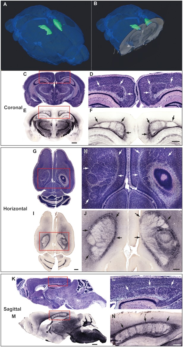
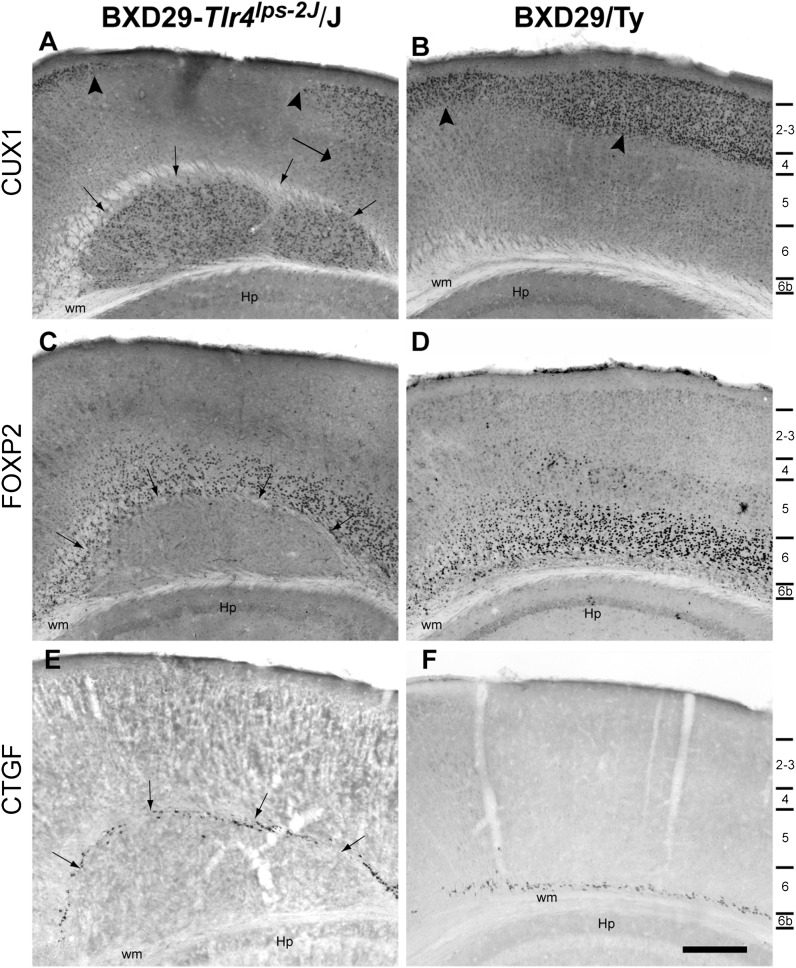
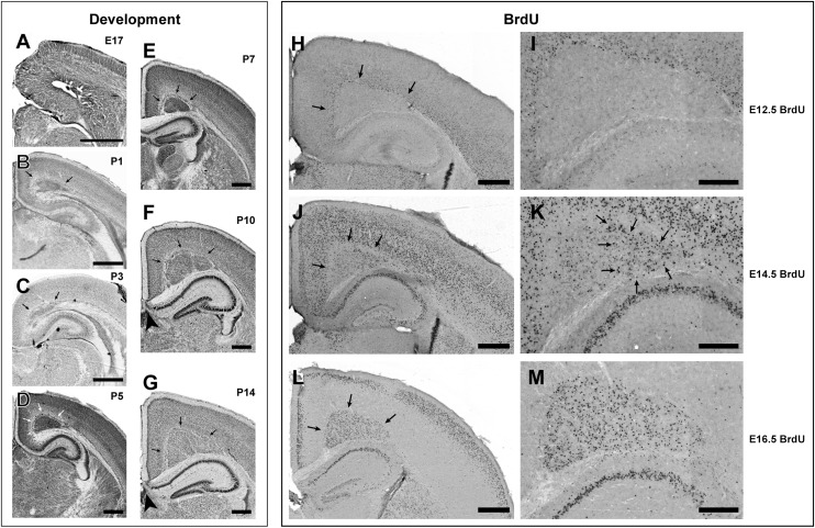
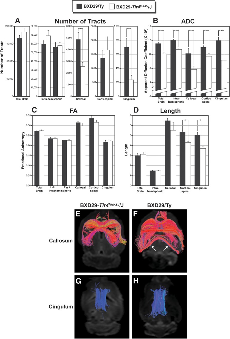
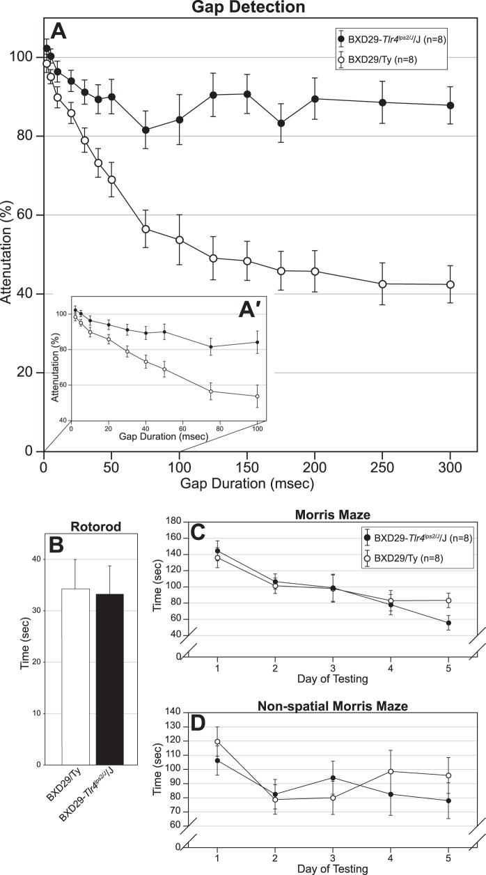
Cognition and behavior depend on the precise placement and interconnection of complex ensembles of neurons in cerebral cortex. Mutations that disrupt migration of immature neurons from the ventricular zone to the cortical plate have provided major insight into mechanisms of brain development and disease. We have discovered a new and highly penetrant spontaneous mutation that leads to large nodular bilateral subcortical heterotopias with partial callosal agenesis. The mutant phenotype was first detected in a colony of fully inbred BXD29 mice already known to harbor a mutation in Tlr4. Neurons confined to the heterotopias are mainly born in midgestation to late gestation and would normally have migrated into layers 2-4 of overlying neocortex. Callosal cross-sectional area and fiber number are reduced up to 50% compared with coisogenic wildtype BXD29 substrain controls. Mutants have a pronounced and highly selective defect in rapid auditory processing. The segregation pattern of the mutant phenotype is most consistent with a two-locus autosomal recessive model, and selective genotyping definitively rules out the Tlr4 mutation as a cause. The discovery of a novel mutation with strong pleiotropic anatomical and behavioral effects provides an important new resource for dissecting molecular mechanisms and functional consequences of errors of neuronal migration.


Figures

Figure 1.
Figure 1.
Bilateral midline neocortical nodular heterotopias in BXD29-Tlr4lps-2J/J mutant mouse neocortex. (A,B) 3D reconstruction of bilateral midline nodular heterotopia reconstructed from high-resolution magnetic resonance imaging (courtesy of G.A. Johnson, Duke Center for In Vivo Microscopy). (C) Nissl stain of section in the coronal plane with bilateral midline neocortical nodular heterotopia. Red box indicates area of enlargement in panel D. (D) Enlarged area from panel B. Arrows denote bilateral midline nodular heterotopia. (E) Section adjacent to panel C immunohistochemically stained for MBP. Red box indicates area of enlargement in panel F. (F) Enlarged area from panel E. Arrows denote bilateral midline nodular heterotopia. Bar in E for C,E = 1 mm. Bar in F for D,F = 500 μm. (G) Nissl stain of section in the horizontal plane with bilateral midline neocortical nodular heterotopia. Red box indicates area of enlargement in panel H. (H) Arrows denote bilateral midline nodular heterotopia. (I) Section adjacent to panel G immunohistochemically stained for MBP. Red box indicates area of enlargement in panel J. (J) Enlarged area from panel I. Arrows denote heterotopia. Bar in I for G,I = 1 mm. Bar in J for H,J = 500 μm. (K) Nissl stain of section in the sagittal plane with bilateral midline neocortical nodular heterotopia. Red box indicates area of enlargement in panel L. (L) Arrows denote bilateral midline nodular heterotopia. (M) Section adjacent to panel K immunohistochemically stained for MBP. Red box indicates area of enlargement in panel M. (N) Enlarged area from panel M. Arrows denote heterotopia. Bar in M for K,M = 1 mm. Bar in N for L,N = 500 μm.
Figure 2.
Figure 2.
Photomontages of MBP-stained midsagittal sections from BXD29-Tlr4lps-2J/J mutant (A) and BXD-29/Ty wildtype (B) mice illustrating partial agenesis of the corpus callosum. Boxes indicate area of enlargement in (C,D). Coronal sections from mutant (E) and wildtype (F) mice at 300 μm intervals indicating that agenesis of the caudal portion of the corpus callosum coincides with the onset of the malformation. Bars for A,B,E,F = 1 mm. Bar for C,D = 250 μm.
Figure 3.
Figure 3.
Laminar markers in BXD29-Tlr4lps-2J/J mutant (A,C,E) and BXD29/Ty wildtype (B,D,F) mice. Photomicrograph of CUX1 immunoreactivity in heterotopia in mutant (A) and wildtype (B) mouse. Small arrows in panel A denote border of nodular heterotopia, which contains large numbers of CUX1 immunopositive neurons. Arrowheads in A highlight region of decreased density of CUX1 immunopositive neurons when compared to homologous region in WT neocortex in B. Large arrow denotes numbers of CUX1-positive neurons in lower cortical laminae, denoted on right side of figure. (C,D) Low power photomicrograph of section from mutant (C) and wildtype mice (D) immunohistochemically stained for FOXP2. There are immunopositive neurons superior to the border of the heterotopia. (E,F) Low power photomicrograph of CTGF immunoreactive neurons in mutant (E) and wildtype mice (F). CTGF neurons are located superior to the nodular heterotopia, and there are no CTGF neurons in the heterotopia itself (E). Hp = hippocampus, wm = white matter. Bar = 250 μm.
Figure 4.
Figure 4.
Development of bilateral subcortical midline heterotopias. Nissl-stained photomicrographs illustrating progression of heterotopia at E17 (A), P1 (B), P3 (C), P5 (D), P7 (E), P10 (F), and P14 (G). The first direct evidence of heterotopia formation appears at P1 (arrows in B). The density of neurons within the heterotopia decreases over time, as the volume of the heterotopia expands in concert with the expansion of the neocortex. Agenesis of the caudal portion of the corpus callosum can be seen in panels F and G (arrowheads). Panels H–M illustrate the results of BrdU experiments. (H) Low power micrograph illustrating the disposition of BrdU+ neurons in mutant mice following E12.5 injection. There are virtually no neurons in the heterotopia. (I) High power photomicrograph of panel H illustrating BrdU+ neurons in layer 6 directly dorsal to the heterotopia, but no neurons within. (J) Low power micrograph illustrating the disposition of BrdU+ neurons in mutant mice following E14.5 injection. There are relatively few densely labeled BrdU + neurons in the lateral-most regions of the heterotopia. (K) High power photomicrograph of panel J illustrating clusters of BrdU+ neurons in the lateral-most portion of the heterotopia (arrows). (L) Low power photomicrograph illustrating the disposition of BrdU+ neurons in mutant mice following E16.5 injection. There are large numbers of densely labeled BrdU+ neurons in the heterotopia and in layers 2–3 of the neocortex. (M) High power photomicrograph of panel L illustrating clusters of BrdU+ neurons throughout the heterotopia but with a somewhat sparser distribution in the lateral portion of the heterotopia.
Figure 5.
Figure 5.
High angular resolution diffusion imaging (HARDI) tractography of the brains of BXD29-Tlr4lps-2J/J mutant and BXD29/Ty wildtype mice. Quantitative results of tract number (A), ADC (B), FA (C), and length (D) from total brain, intrahemispheric (right and left), callosal, corticospinal, and cingulum of wildtype (black bars) and mutant (white bars). (A) There is a significant decrease in the number of tractography pathways passing through the corpus callosum of mutant mice when compared to the wildtype. In addition, there is a decrease in the number of cingulum tractography pathways in the mutant mice. There were no significant differences in the number of tracts in any of the other tractography pathways. (B) There is a significant decrease in ADC in mutant compared with wildtype mice across all tract categories. (C) There is no significant difference in FA among the tract categories. (D) The length of corticospinal tracts and cingulum are decreased in the mutant as compared to wildtype. There are no significant differences in the length of fiber tracts in any of the other tract measures. (E,F) Visualization of tractography pathways passing through the corpus callosum of mutant (E) and wildtype (F) mice. There are fewer callosal tracts in the mutant as compared to the wildtype, and there is a lack of interhemispheric tracts in the caudal regions of the forebrain (arrows). (G,H). Visualization of cingulum fiber tracts in the left and right hemispheres of mutant (G) and wildtype (H) mice. There are fewer and shorter fibers in the mutant cingulum as compared to the wildtype.
Figure 6.
Figure 6.
Behavioral tests of BXD29-Tlr4lps-2J /J mutant and BXD29/Ty wildtype strain. (A) SG 0–300 ms and SG 0–100 ms data are combined; see (A') for SG 0–100 ms data only. Mutant mice are significantly impaired in rapid auditory processing as assessed by startle reduction in a gap detection task. There is no difference (and no attenuation between mutant and wildtype mice at the shortest gap durations [50–150 ms and 2–10 ms] for the SG 0–300 and SG 0–100 ms task, respectively see A and A′, respectively), but the mutant significantly attenuates at the 300 ms gap durations for the SG 0–300 ms task and at the 100 ms gap for the SG 0–100 ms task. There are no differences between the strains on rotarod (B), Morris maze (C), or nonspatial Morris maze (D).

References

    1. Andrade DM. Genetic basis in epilepsies caused by malformations of cortical development and in those with structurally normal brain. Hum Genet. 2009;126:173–193. - PubMed
    1. Bai J, Ramos RL, Ackman JB, Thomas AM, Lee RV, LoTurco JJ. RNAi reveals doublecortin is required for radial migration in rat neocortex. Nat Neurosci. 2003;6:1277–1283. - PubMed
    1. Bilasy SE, Satoh T, Ueda S, Wei P, Kanemura H, Aiba A, Terashima T, Kataoka T. Dorsal telencephalon-specific RA-GEF-1 knockout mice develop heterotopic cortical mass and commissural fiber defect. Eur J Neurosci. 2009;29:1994–2008. - PubMed
    1. Caviness VS, Jr, Frost DO. Thalamocortical projections in the reeler mutant mouse. J Comp Neurol. 1983;219:182–202. - PubMed
    1. Caviness VS, Jr, So DK, Sidman RL. The hybrid reeler mouse. J Hered. 1972;63:241–246. - PubMed

Publication types

MeSH terms

Supplementary concepts