. 2012 Apr 24;12(10):1813-22.
doi: 10.1039/c2lc21202a. Epub 2012 Mar 28.

A programmable microfluidic cell array for combinatorial drug screening

Affiliations

A programmable microfluidic cell array for combinatorial drug screening

Jeongyun Kim et al. Lab Chip. .

Abstract

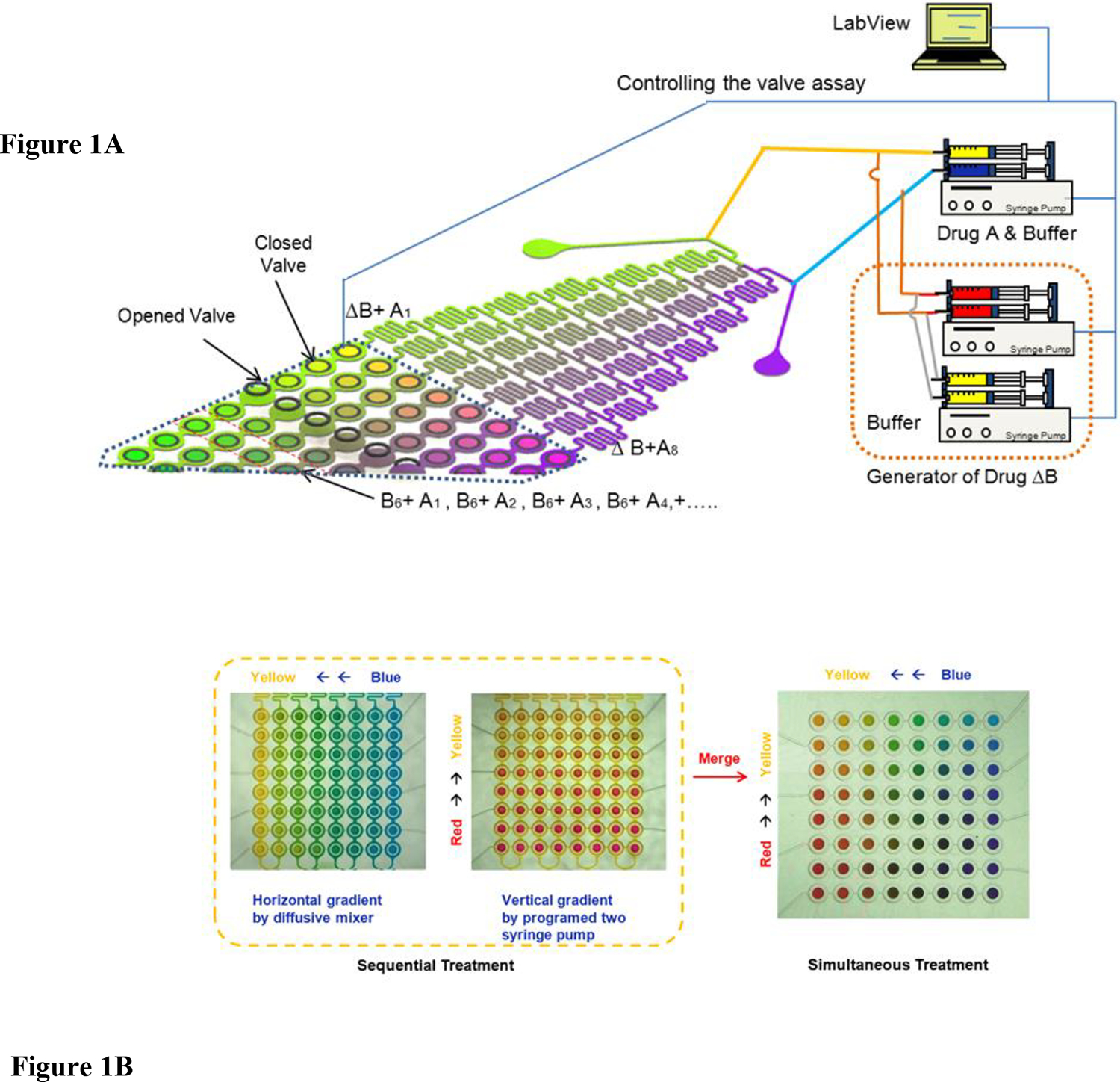
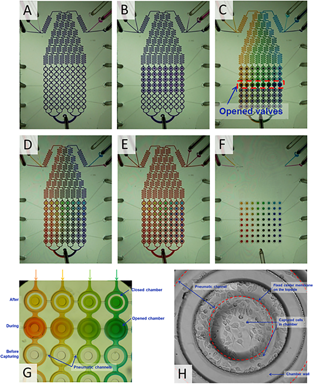
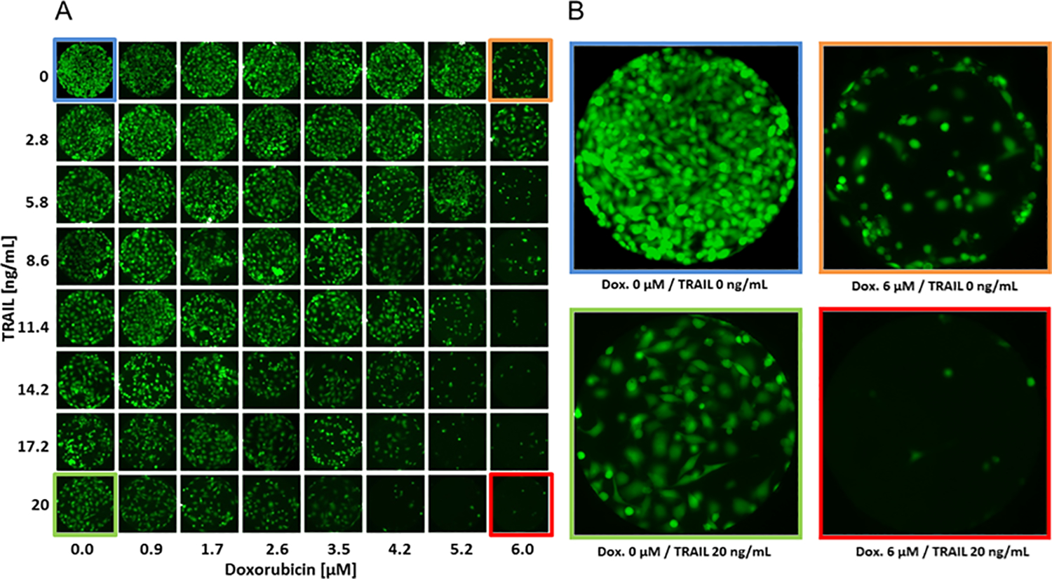
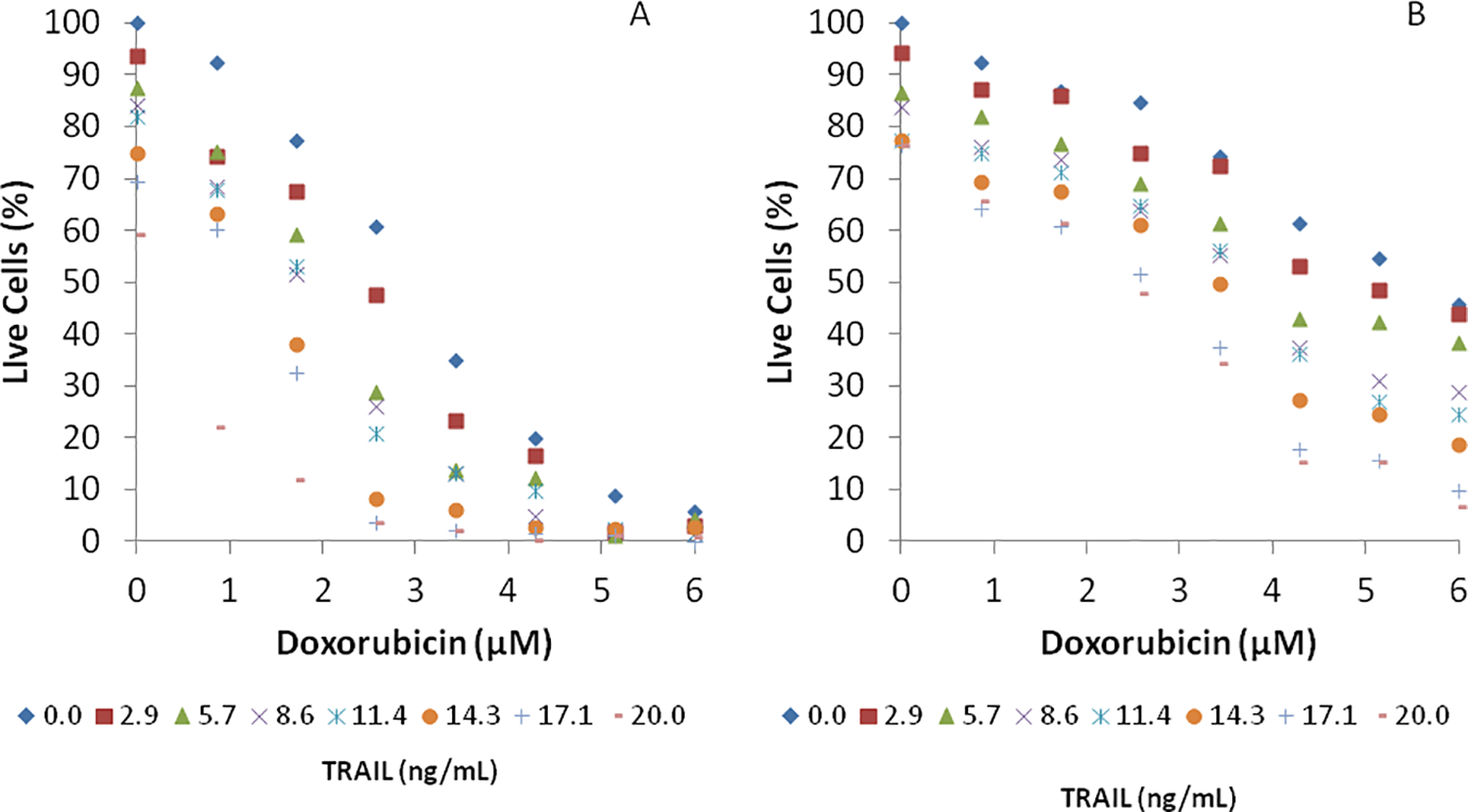
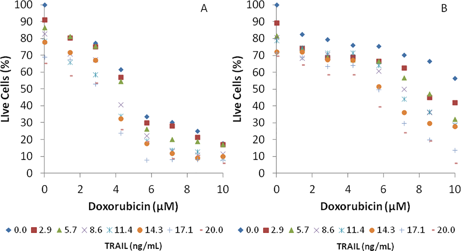
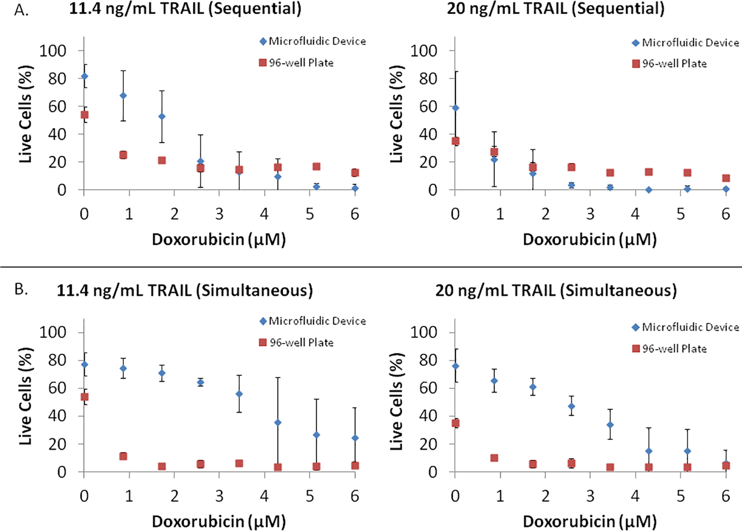
We describe the development of a fully automatic and programmable microfluidic cell culture array that integrates on-chip generation of drug concentrations and pair-wise combinations with parallel culture of cells for drug candidate screening applications. The device has 64 individually addressable cell culture chambers in which cells can be cultured and exposed either sequentially or simultaneously to 64 pair-wise concentration combinations of two drugs. For sequential exposure, a simple microfluidic diffusive mixer is used to generate different concentrations of drugs from two inputs. For generation of 64 pair-wise combinations from two drug inputs, a novel time dependent variable concentration scheme is used in conjunction with the simple diffusive mixer to generate the desired combinations without the need for complex multi-layer structures or continuous medium perfusion. The generation of drug combinations and exposure to specific cell culture chambers are controlled using a LabVIEW interface capable of automatically running a multi-day drug screening experiment. Our cell array does not require continuous perfusion for keeping cells exposed to concentration gradients, minimizing the amount of drug used per experiment, and cells cultured in the chamber are not exposed to significant shear stress continuously. The utility of this platform is demonstrated for inducing loss of viability of PC3 prostate cancer cells using combinations of either doxorubicin or mitoxantrone with TRAIL (TNF-alpha Related Apoptosis Inducing Ligand) either in a sequential or simultaneous format. Our results demonstrate that the device can capture the synergy between different sensitizer drugs and TRAIL and demonstrate the potential of the microfluidic cell array for screening and optimizing combinatorial drug treatments for cancer therapy.


Figures

Figure 1.
Figure 1.. Schematic of the microfluidic array.
(A) Different concentrations of Drugs A and B are generated in the diffusive gradient mixer, and used, either sequentially or in combination, to perfuse cells cultured in downstream microchambers. The mixing operation for generating different drug combinations and the opening and closing of valves for perfusing cells in the microchambers are controlled though a LabView interface. (B) Depiction of the range of concentrations that can be generated for sequential and simultaneous treatment using color dyes. In the left panel, yellow and blue color dye solutions (representing the minimum and maximum concentrations of a drug) are mixed to generate eight outlet concentrations (“horizontal gradient” of colors between yellow and blue), and represent the gradient used in sequential exposure experiments. In the middle panel, yellow and red streams (representing the minimum and maximum concentrations of the second drug) are mixed together to generate a “vertical gradient” of colors between yellow and red. Merging the two color gradients (vertical direction concentration gradient: yellow to blue; horizontal direction concentration gradient: yellow to red) yields an array of pair-wise combinations, and represents the gradient used in simultaneous exposure experiments (right panel).
Figure 2.
Figure 2.. Sequence of steps in the operation of the combinatorial array.
(A) Isolation of cell culture chambers shown by flowing violet dye around it. (B) Trapping of violet dye in the cell culture chambers. (C) Sequential trapping of different drug concentrations (represented by different colors) in columns of cell culture chambers. (D) Array of colors trapped in the chambers, with each color representing a pair-wise combination of two drugs. (E) No mixing occurs between the color dye present in the cell culture chamber and the color dye flowing outside. (F) Color dyes combinations trapped in the chambers without any liquid flowing around it. (G) Sequential operation of pneumatically controlled trapping system. Three sets of chambers are shown: empty chambers prior to trapping of color dye solution, chambers during trapping, and chambers after trapping of solution. (H) Representative cell culture chamber with PC3 cells trapped and grown for 24 h.
Figure 3.
Figure 3.. Cell culture in the combinatorial array.
(A) Representative fluorescent micrographs of PC3 cells cultured in an array for 24 h. (B) Micrographs of cells in the four corners of the array after sequential exposure to Doxorubicin and TRAIL (top left: no Doxorubicin, no TRAIL; top right: Doxorubicin, no TRAIL; bottom left: No Doxorubicin, TRAIL; bottom right: Doxorubicin, TRAIL).
Figure 4.
Figure 4.. PC3 cell viability after sequential exposure to Doxorubicin and TRAIL.
PC3 cells were initially exposed to (A) eight concentrations (0.0, 0.9, 1.7, 2.6, 3.5, 4.2, 5.2, and 6.0 μM) of Doxorubicin or (B) eight concentrations (0.0, 1.4, 2.9, 4.3, 5.7, 7.1, 8.6, and 10 μM) of Mitoxantrone for 24 h, followed by exposure to eight concentrations (0.0, 2.9, 5.7, 8.6, 11.4, 14.3, 17.1, and 20.0 ng /mL) of TRAIL for 24 h. Data shown are average of three independent experiments.
Figure 5.
Figure 5.. PC3 cell viability after simultaneous exposure to pairwise combinations.
PC3 cells were exposed to 64 pair-wise combinations of (A) eight concentrations (0.0, 0.9, 1.7, 2.6, 3.5, 4.2, 5.2, and 6.0 μM) Doxorubicin and eight concentrations (0.0, 2.9, 5.7, 8.6, 11.4, 14.3, 17.1, and 20.0 ng /mL) of TRAIL or (B) eight concentrations (0.0, 1.4, 2.9, 4.3, 5.7, 7.1, 8.6, and 10 μM) of Mitoxantrone and eight concentrations (0.0, 2.9, 5.7, 8.6, 11.4, 14.3, 17.1, and 20.0 ng /mL) of TRAIL for 24 h. Data shown are average of three independent experiments.
Figure 6.
Figure 6.. Comparison of PC3 viability in microfluidic array and tissue culture plates.
PC3 cell viability in response to (A) sequential exposure to eight concentrations (0.0, 0.9, 1.7, 2.6, 3.5, 4.2, 5.2, and 6.0 μM) of Doxorubicin for 24 h, followed by exposure to 11.4 ng/mL or 20 ng/mL TRAIL for 24 h, and (B) simultaneous exposure to eight concentrations (0.0, 0.9, 1.7, 2.6, 3.5, 4.2, 5.2, and 6.0 μM) of Doxorubicin and 11.4 ng/mL or 20 ng/mL TRAIL for 24 h, was compared between the microfluidic array and tissue culture plates. Data shown are average of three independent experiments and one standard deviation.

References

    1. Beltran H, Beer TM, Carducci MA, de Bono J, Gleave M, Hussain M, Kelly WK, Saad F, Sternberg C, Tagawa ST and Tannock IF, Eur Urol, 2011, 60, 279–290. - PubMed
    1. Zhang L and Fang B, Cancer Gene Ther, 2005, 12, 228–237. - PubMed
    1. Sheikh MS and Fornace AJ Jr, J Cell Physiol, 2000, 182, 171–181. - PubMed
    1. Bouralexis S, Findlay DM and Evdokiou A, Apoptosis, 2005, 10, 35–51. - PubMed
    1. Kelley SK and Ashkenazi A, Curr Opin Pharmacol, 2004, 4, 333–339. - PubMed

Publication types

MeSH terms

Substances

LinkOut - more resources