. 2012 Apr;7(2):024104.
doi: 10.1088/1748-6041/7/2/024104. Epub 2012 Mar 29.

Injectable biomaterials for adipose tissue engineering

Affiliations

Injectable biomaterials for adipose tissue engineering

D A Young et al. Biomed Mater. 2012 Apr.

Abstract

Adipose tissue engineering has recently gained significant attention from materials scientists as a result of the exponential growth of soft tissue filler procedures being performed within the clinic. While several injectable materials are currently being marketed for filling subcutaneous voids, they often face limited longevity due to rapid resorption. Their inability to encourage natural adipose formation or ingrowth necessitates repeated injections for a prolonged effect and thus classifies them as temporary fillers. As a result, a significant need for injectable materials that not only act as fillers but also promote in vivo adipogenesis is beginning to be realized. This paper will discuss the advantages and disadvantages of commercially available soft tissue fillers. It will then summarize the current state of research using injectable synthetic materials, biopolymers and extracellular matrix-derived materials for adipose tissue engineering. Furthermore, the successful attributes observed across each of these materials will be outlined along with a discussion of the current difficulties and future directions for adipose tissue engineering.


Figures

Figure 1
Figure 1. Adipose Tissue and Preadipocytes
Histology of adipose tissue, visualized with H&E staining, reveals its characteristic architecture (A). The “voids” seen in this image are the location of lipid-filled vacuoles that have been washed away as a result of the staining reagents. Adipose-derived stem cells can be isolated from adipose tissue and differentiated in vitro into adipocytes that collect lipid filled vacuoles, stained here with Oil Red O (B). Scale bars = 100 μm.
Figure 2
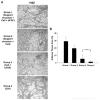
Figure 2. Fibrin Injections for Adipose Tissue Engineering
Cho et al showed improved adipogenesis at 6 weeks when a PGA support structure was implanted subcutaneously to protect fibrin + preadipocyte injections from tissue constriction (Groups 1 & 2). Addition of bFGF to fibrin + preadipocyte injections further enhanced lipid accumulation (Groups 1 & 3). Group 4 also shows the relative inability of fibrin + preadipocyte injections to produce fatty tissue on their own. Scale bar = 200 μm (A). The relative amount of adipose tissue present was quantified using an Oil Red O stain to verify that the support structures significantly increased adipogenesis (B). Reprinted in part with permission from [37].
Figure 3
Figure 3. Injection of Photoactivated PEG-HA Hydrogel
Hillel et al were able to create an in situ crosslinking injectable filler by combining hyaluronic acid (HA) with PEG-diacrylate and the photoinitiator, eosin Y. This mixture of components was injected transdermally (A) and could be massaged into a desired shape (B). The material was then crosslinked by shining an array of light emitting diodes (LEDs) emitting light at a wavelength of 520 nm (C). This light was shown to penetrate up to tissue depths of 4 mm, and a 2 minute exposure time was sufficient to activate the eosin Y and photocrosslink the composite implant. Reprinted with permission from [52].
Figure 4
Figure 4. bFGF Required for Matrigel-induced Adipogenesis in vivo
Subcutaneous injections of Matrigel and 1 μg/mL bFGF were performed in mice and H&E stained sections were analyzed over a 10-week period by Kawaguchi et al [57]. After 1 week, neovascularization could be seen in the injection region (A), and by 2 weeks fibroblast-like cells had invaded and differentiated into adipocytes (B). At 5 weeks, significant adipocyte formation and maturation was seen in the injection region (C), which persisted through the entire 10-week duration of the study (D). However, when bFGF was not included in the Matrigel injections, limited adipogenesis was seen (E). Copyright 1998, National Academy of Sciences, USA.
Figure 5
Figure 5. Lipoaspirate-derived Hydrogels
Human lipoaspirate was decellularized and delipidized to isolate adipose extracellular matrix components (A). This powder was subsequently reduced to liquid form via enzymatic digestion (B). This formulation would then self-assemble into a soft hydrogel in vitro when brought to physiologic temperature (C). Subcutaneous injections of the liquid matrix into nude mice would also self-assemble within 15 minutes (D). Arrows indicate subcutaneous hydrogels of injected adipose matrix.

References

    1. Wang P, Mariman E, Keijer J, Bouwman F, Noben J-P, Robben J, Renes J. Profiling of the secreted proteins during 3t3-l1 adipocyte differentiation leads to the identification of novel adipokines. Cell Mol Life Sci. 2004;61:2405–17. - PMC - PubMed
    1. Zuk P, Zhu M, Mizuno H, Huang J, Futrell J, Katz A, Benhaim P, Lorenz H, Hedrick M. Multilineage cells from human adipose tissue: Implications for cell-based therapies. Tissue Engineering. 2001;7:211–28. - PubMed
    1. Prichard HL, Reichert W, Klitzman B. Ifats collection: Adipose-derived stromal cells improve the foreign body response. Stem Cells. 2008;26:2691–5. - PMC - PubMed
    1. Gomillion CT, Burg KJL. Stem cells and adipose tissue engineering. Biomaterials. 2006;27:6052–63. - PubMed
    1. Flynn L, Woodhouse K. Adipose tissue engineering with cells in engineered matrices. Organogenesis. 2008;4:228. - PMC - PubMed

Publication types