. 2012 Jul;40(13):6353-66.
doi: 10.1093/nar/gks263. Epub 2012 Mar 28.

Transient structure and dynamics in the disordered c-Myc transactivation domain affect Bin1 binding

Affiliations

Transient structure and dynamics in the disordered c-Myc transactivation domain affect Bin1 binding

Cecilia Andresen et al. Nucleic Acids Res. 2012 Jul.

Abstract

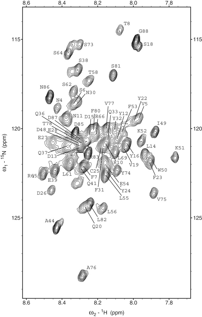
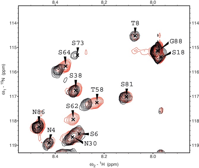
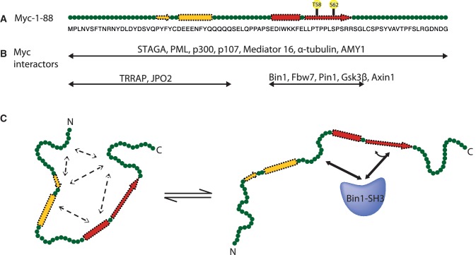
The crucial role of Myc as an oncoprotein and as a key regulator of cell growth makes it essential to understand the molecular basis of Myc function. The N-terminal region of c-Myc coordinates a wealth of protein interactions involved in transformation, differentiation and apoptosis. We have characterized in detail the intrinsically disordered properties of Myc-1-88, where hierarchical phosphorylation of S62 and T58 regulates activation and destruction of the Myc protein. By nuclear magnetic resonance (NMR) chemical shift analysis, relaxation measurements and NOE analysis, we show that although Myc occupies a very heterogeneous conformational space, we find transiently structured regions in residues 22-33 and in the Myc homology box I (MBI; residues 45-65); both these regions are conserved in other members of the Myc family. Binding of Bin1 to Myc-1-88 as assayed by NMR and surface plasmon resonance (SPR) revealed primary binding to the S62 region in a dynamically disordered and multivalent complex, accompanied by population shifts leading to altered intramolecular conformational dynamics. These findings expand the increasingly recognized concept of intrinsically disordered regions mediating transient interactions to Myc, a key transcriptional regulator of major medical importance, and have important implications for further understanding its multifaceted role in gene regulation.


Figures

Figure 1.
Figure 1.
Domain structure of c-Myc, indicating conserved Myc homology boxes MBI, MBII, MBIIIa, MBIIIb and MBIV (1). The basic region (BR) N-terminal to the HLHLZ binds DNA in a heterodimeric complex with Max (5). The Myc fragments 1–88 studied in this work is indicated together with an expansion of MBI and phosphorylation sites T58 and S62 located therein.
Figure 2.
Figure 2.
15N-HSQC spectrum of Myc-1–88 recorded at 15°C with overlaid assignments. The glycine region (δ 15N < 112 ppm) has been omitted for clarity but contains peaks corresponding to G84 and G68.
Figure 3.
Figure 3.
Identification of transient structure in Myc-1–88 by NMR. (A) SSP of Myc-1–88 based on experimental data. A region with values close to one indicates a fully formed α-helix whereas values close to minus one and zero are indicative of fully formed β-strand or no preferential secondary structure, respectively (47). (B) Sequence conservation in Myc-1–88 described on a normalized c-score scale as described in ‘Materials and methods’ section: high (0.8–1.0, star) intermediate (0.6–0.8, dot) and low (0–0.6, unlabeled). (C) Graphical representation of transient structures based on combined SSP and NOE evaluation. (D) Schematic representation of characteristic secondary structure NOEs as identified in 15N-NOESY-HSQC spectra. The dNN(i, i + 1) NOEs are classified as strong, intermediate or weak by the intensity of the bar, while other NOEs are indicated as present irrespective of magnitude. Light gray indicates partial overlap whereas completely overlapping NOEs are excluded.
Figure 4.
Figure 4.
SPR measurements suggest multivalent binding of Myc-1–88 to Bin1–SH3. Myc-1–88 was immobilized and Bin1–SH3 injected over the surface. (A) Kinetic experiments. Overlaid sensorgrams show experimental data (solid lines) and simultaneously fitted functions using a parallel reaction model with two binding sites (dashed lines). A 1:1 Langmuir model fits very poorly as indicated to the middle response (gray line); no Langmuir fit could be made to all curves simultaneously. The concentration series includes 111.0, 55.5, 27.7, 13.9 and 6.9 µM of Bin1–SH3. (B) Steady-state binding experiments of Bin1–SH3 to Myc-1–88. Based on the data points at lower concentrations a plateau in binding would be expected at higher concentration, but instead the response increases further in both kinetic and steady-state experiments, thus indicating that Myc-1–88 may display one or more additional binding sites for Bin1–SH3 with lower affinity.
Figure 5.
Figure 5.
Selected HSQC region showing superimposed spectra of Myc-1–88 in the free state (red, labeled) and in a 1:1.5 complex with Bin1–SH3 (black, unlabeled). In the bound state, the resonance for S62 is broadened beyond detection, thus no peak for S62 is observed in the bound state in the selected region or elsewhere.
Figure 6.
Figure 6.
NMR analysis of Myc-1–88 in the absence and presence of Bin1–SH3. Omitted histogram bars correspond to missing or overlapped residues and prolines. (A) Myc-1–88 CSPs at a Myc-1–88:Bin1–SH3 ratio of 1:1.5. The cut-off value for significant CSPs is shown as a dashed line and calculated as described in ‘Materials and methods’ section. (B) Ratios of peak intensities with and without Bin1, derived from HNCO experiments at a Myc-1–88:Bin1–SH3 ratio of 1:1.5. In the absence of interactions, or if interactions are the same in free and bound forms, the intensity ratio would be 1 (gray line). Gain/loss of interactions in the bound state lead to decreased/increased peak intensity ratio, respectively.
Figure 7.
Figure 7.
15N Relaxation parameters of free Myc-1–88 (open circles) and Myc-1–88 bound to Bin1–SH3 in a 1:1.5 ratio (filled circles). (A) {1H}-15N-NOE. (B) R1 relaxation rate constants. (C) R2 relaxation rate constants.
Figure 8.
Figure 8.
Biophysical and biological functionalities in Myc-1–88. (A) Myc-1–88 is intrinsically disordered (green), with transient secondary propensity in residues 23–33 (yellow) and 48-65 (red); phosphorylation sites T58 and S62 are indicated. (B) Identified interactors to Myc-1–88 can be classified in three groups as indicated by double arrows: those mapped by deletion mutants covering entire Myc1–88, those identified to interact with MBI by deletion mapping or where interaction is regulated by phosphorylation to T58 and/or S62, and those that bind the N-terminal region including the first transiently structured region. (C) In the free state, Myc-1–88 shows transient and fluctuating interactions (dashed arrows) involving secondary structure elements and hydrophobic clusters, suggesting the presence of condensed disordered states in the conformational ensemble (only one of many such possible states is shown). By multivalent binding to Bin1 binding sites at P59–P63 and P42–P45 (solid arrows) and possible wobbling, or sliding, to the less optimal P57–P60 site (curved arrow), Bin1 sterically hinders interactions present in the free state.

References

    1. Meyer N, Penn LZ. Reflecting on 25 years with MYC. Nat. Rev. Cancer. 2008;8:976–990. - PubMed
    1. Eilers M, Eisenman RN. Myc’s broad reach. Genes Dev. 2008;22:2755–2766. - PMC - PubMed
    1. Agrawal P, Yu K, Salomon AR, Sedivy JM. Proteomic profiling of Myc-associated proteins. Cell Cycle. 2010;9:4908–4921. - PMC - PubMed
    1. Kim J, Woo AJ, Chu J, Snow JW, Fujiwara Y, Kim CG, Cantor AB, Orkin SH. A Myc network accounts for similarities between embryonic stem and cancer cell transcription programs. Cell. 2010;143:313–324. - PMC - PubMed
    1. Kalkat M, Wasylishen AR, Kim SS, Penn L. More than MAX: discovering the Myc interactome. Cell Cycle. 2011;10:374–379. - PubMed

Publication types

MeSH terms