. 2012 Jul;40(13):e101.
doi: 10.1093/nar/gks284. Epub 2012 Mar 28.

Fluorescence-based incision assay for human XPF-ERCC1 activity identifies important elements of DNA junction recognition

Affiliations

Fluorescence-based incision assay for human XPF-ERCC1 activity identifies important elements of DNA junction recognition

Maureen Bowles et al. Nucleic Acids Res. 2012 Jul.

Abstract

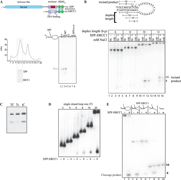
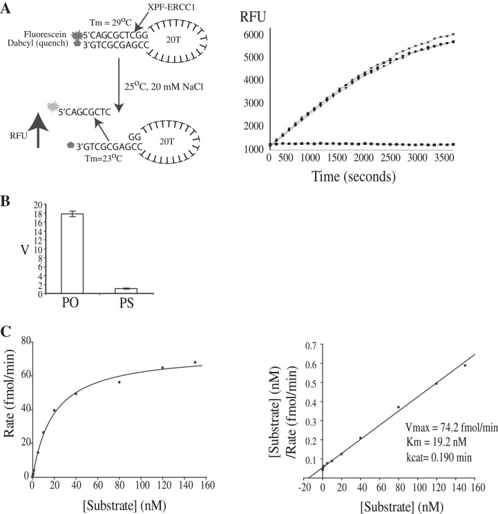
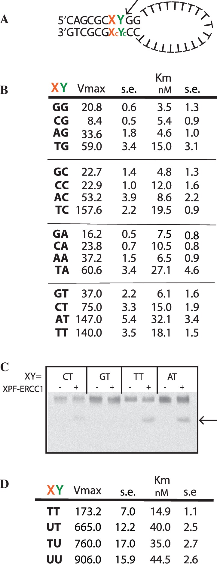
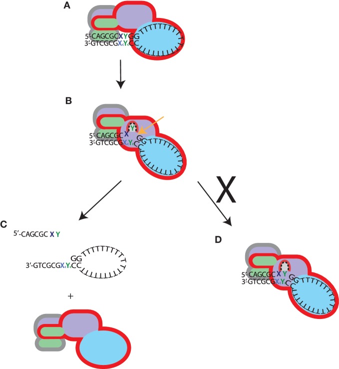
The structure-specific endonuclease activity of the human XPF-ERCC1 complex is essential for a number of DNA processing mechanisms that help to maintain genomic integrity. XPF-ERCC1 cleaves DNA structures such as stem-loops, bubbles or flaps in one strand of a duplex where there is at least one downstream single strand. Here, we define the minimal substrate requirements for cleavage of stem-loop substrates allowing us to develop a real-time fluorescence-based assay to measure endonuclease activity. Using this assay, we show that changes in the sequence of the duplex upstream of the incision site results in up to 100-fold variation in cleavage rate of a stem-loop substrate by XPF-ERCC1. XPF-ERCC1 has a preference for cleaving the phosphodiester bond positioned on the 3'-side of a T or a U, which is flanked by an upstream T or U suggesting that a T/U pocket may exist within the catalytic domain. In addition to an endonuclease domain and tandem helix-hairpin-helix domains, XPF has a divergent and inactive DEAH helicase-like domain (HLD). We show that deletion of HLD eliminates endonuclease activity and demonstrate that purified recombinant XPF-HLD shows a preference for binding stem-loop structures over single strand or duplex alone, suggesting a role for the HLD in initial structure recognition. Together our data describe features of XPF-ERCC1 and an accepted model substrate that are important for recognition and efficient incision activity.


Figures

Figure 1.
Figure 1.
Minimum substrate requirements for incision activity. (A) Top: domain organization of the human XPF–ERCC1 complex; bottom left panel: gel-filtration profile of XPF–ERCC1 expressed in E. coli after purification on nickel-agarose, and heparin–Sepharose. XPF–ERCC1 eluted where indicated. Fractions 7–8 is the void and the peak at 11 ml corresponded to a MW of ∼250 kDa, and 13 ml to 35 kDa. Inset shows 12% SDS–PAGE gel of fraction 12 stained with Coomassie blue. XPF migrates at 115 kDa and ERCC1 at 36 kDa. Bottom right panel: sequencing gel showing cleavage of 200 fmol stem–loop substrate by wild-type XPF–ERCC1 and XPF-D731N–ERCC1 mutant. Lanes 1 and 5 contained protein buffer from each preparation of protein, lanes 2–4 contained 12, 24 and 48 nM XPF–ERCC1, respectively. Lanes 6–8 contained 12, 24 and 48 nM XPF-D731N–ERCC1. (B) Sequencing gel showing cleavage of stem–loops with either 6 8, 10 or 12 bp duplex stems. Wells contained protein buffer (−) or 42.6 nM XPF–ERCC1 (+) in buffer containing either 50 or 100 mM NaCl as indicated. Cleavage of the 12-bp duplex yields products of 10 and 8 bp, and the 10-bp duplex is cleaved at one major site yielding an 8-bp product. Dual uncleaved bands in lanes 1–8 are full-length (FL) and FL-1 nucleotide oligonucleotides produced during synthesis of the shorter 6 and 8 bp substrates. (C) Separation of products of cleavage reaction on a 10% denaturing polyacrylamide gel. Stem–loops (100 nM) labelled 5′ with Cy-5 with a phosphorothioate bond replacing the phosphodiester bond 3′ to the base represented in lower case in the sequences 5′-CAGCGCTCGG-3′ (TC), 5′-CAGCGCTcGG-3′ (Tc), and 5′-CAGCGCtCGG-3′(tC) in the top strand of the duplex were incubated with 42.6 nM XPF-ERCC1 for 1 hour. (D) Ten percent sequencing gel showing incision reactions containing stem–loops with single-strand loops of increasing T loop size as indicated. An amount of 400 fmol labelled stem–loop substrates were incubated with buffer only (−) or 42.6 nM XPF–ERCC1 (+). (E) Sequencing gel showing incisions made by XPF–ERCC1 on stem–loop, splayed arm, 3′-overhangs, 5′-overhangs and duplex. The sequence of the duplex portion of the structure is 5′-CAGCGCTCGG-3′/5′CCGAGCGCTG-3′ and single-strand regions are 20T for stem–loop and 10T for splayed arms/overhangs. The top strand of each structure was labelled 5′ as indicated and oligonucleotides were annealed and purified at 15°C as described in experimental procedures. The cleavage product is the same eight-base oligo for each structure as indicated in the figure.
Figure 2.
Figure 2.
A microplate fluorescence-based assay to measure XPF–ERCC1 incision activity. (A) Left panel: principle of the fluorescence-based assay. RFU is relative fluorescence units. Incubation of intact stem–loop with XPF–ERCC1 in 0.75 mM MnCl2 and 20 mM NaCl results in the cleaved oligonucleotide diffusing into solution at 25°C. Right panel: increase in fluorescence (RFU) with time. Reactions are shown in triplicate and contained: 3 nM XPF–ERCC1, 100 nM standard stem–loop substrate, 0.75 nM MnCl2, 0.5 mM DTT and 20 mM NaCl2 in 50 mM Tris buffer pH 8 at 25°C. Solid squares: buffer only (B) Graph showing rate of incisions (v) of 62.5 nM stem–loop by 3 nM XPF–ERCC1 where the scissile phosphate contains non-bridging oxygens (PO) or a non-bridging sulphur (PS). Average of three points ± SD. (C) Left panel: Michaelis–Menten plot showing rate versus substrate concentration for full-length XPF–ERCC1. Mean of three points. Right panel: Hanes–Woolf plot.
Figure 3.
Figure 3.
Local sequence preferences at the XPF–ERCC1 incision site on the substrate affects the rate of cleavage. (A) Stem–loop structure showing the positions of substituted bases X and Y in the duplex. Xc and Yc are the complementary bases to X and Y, A is used complementary to U. (B) Kinetic data where X and Y were substituted with each of the four bases. Data are mean of triplicate samples, ±1 standard error. (C) Incisions produced by 4.27 nM XPF–ERCC1 on 5′ 6-FAM-labelled stem–loop substrates with bases at X and Y as indicated. Arrow shows cleavage product. Reactions were incubated for 15 min at 25°C and therefore did not run to completion. (D) Kinetic data where X and Y were substituted with the indicated bases. Data are mean of triplicate samples +/− standard error.
Figure 4.
Figure 4.
XPF-HLD is required for activity and preferentially binds stem–loops. (A) Top; gel-filtration profile of Δ640XPF–ERCC1 after separation on Superose 12 16/60. Line indicates fractions pooled for analysis. Bottom; 12% SDS–PAGE gel of fractions from gel-filtration column stained with Coomassie blue. Lanes show fractions from a Superose 12 16/60 column. Graph shows incisions by wild-type, catalytically impaired and Δ640-XPF–ERCC1 complexes at 50 nM or 1.3 µM, as indicated. (B) Top: 12% SDS–PAGE gel of purified recombinant XPF–HLD stained with Coomassie blue. Top graph: binding of purified XPF–HLD to stem loops with TT and GG substitutions at X and Y (Figure 3). Bottom graph: binding of XPF–HLD to stem loop with CT substitutions at X and Y, a 15-mer single-strand oligonucleotide, and duplex corresponding to the duplex in the stem–loop with a 2T loop, as indicated.
Figure 5.
Figure 5.
Proposed model for stem–loop substrate recognition by XPF–ERCC1. The XPF protomer has a red outline, and ERCC1, grey outline. The domain colouring is as Figure 1: (HhH)2 domains; green, nuclease or nuclease-like domains; mauve and HLD; blue. (A) The XPF–HLD binds to the junction containing single-strand stretches greater than six bases and the hairpins of ERCC1 occupy five bases of the duplex. (B) A domain movement occurs resulting in bending of the DNA, and the nuclease domain of XPF engages the duplex downstream from the ERCC1 (HhH)2 domain. A pocket in the catalytic centre of XPF becomes occupied by the base 5′ to the scissile phosphate, and cleavage occurs. (C) After incision the complex is released from the substrate. (D) Failure of cleavage due to lack of entry of an appropriate base into the binding pocket, results in the complex remaining bound and slow dissociation from the substrate.

References

    1. Friedberg EC, Aguilera A, Gellert M, Hanawalt PC, Hays JB, Lehmann AR, Lindahl T, Lowndes N, Sarasin A, Wood RD. DNA repair: from molecular mechanism to human disease. DNA Repair. 2006;5:986–996. - PubMed
    1. Sijbers AM, de Laat WL, Ariza RR, Biggerstaff M, Wei YF, Moggs JG, Carter KC, Shell BK, Evans E, de Jong MC, et al. Xeroderma pigmentosum group F caused by a defect in a structure-specific DNA repair endonuclease. Cell. 1996;86:811–822. - PubMed
    1. Niedernhofer LJ, Odijk H, Budzowska M, van Drunen E, Maas A, Theil AF, de Wit J, Jaspers NG, Beverloo HB, Hoeijmakers JH, et al. The structure-specific endonuclease Ercc1-Xpf is required to resolve DNA interstrand cross-link-induced double-strand breaks. Mol. Cell Biol. 2004;24:5776–5787. - PMC - PubMed
    1. Sargent RG, Meservy JL, Perkins BD, Kilburn AE, Intody Z, Adair GM, Nairn RS, Wilson JH. Role of the nucleotide excision repair gene ERCC1 in formation of recombination-dependent rearrangements in mammalian cells. Nucleic Acids Res. 2000;28:3771–3778. - PMC - PubMed
    1. Ahmad A, Robinson AR, Duensing A, van Drunen E, Beverloo HB, Weisberg DB, Hasty P, Hoeijmakers JH, Niedernhofer LJ. ERCC1-XPF endonuclease facilitates DNA double-strand break repair. Mol. Cell Biol. 2008;28:5082–5092. - PMC - PubMed

Publication types