. 2012 May 25;287(22):18645-54.
doi: 10.1074/jbc.M112.345736. Epub 2012 Mar 28.

Mechanisms for defining supercoiling set point of DNA gyrase orthologs: II. The shape of the GyrA subunit C-terminal domain (CTD) is not a sole determinant for controlling supercoiling efficiency

Affiliations

Mechanisms for defining supercoiling set point of DNA gyrase orthologs: II. The shape of the GyrA subunit C-terminal domain (CTD) is not a sole determinant for controlling supercoiling efficiency

Elsa M Tretter et al. J Biol Chem. .

Abstract

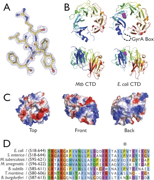
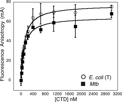
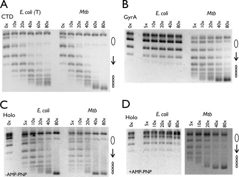
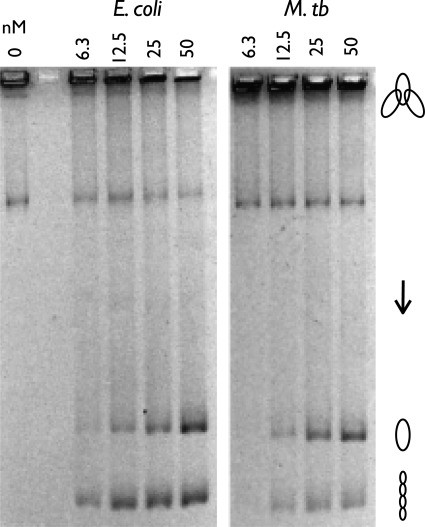
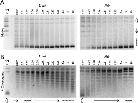
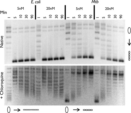
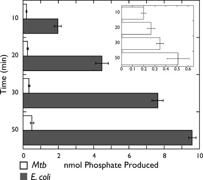
DNA topoisomerases are essential enzymes that can overwind, underwind, and disentangle double-helical DNA segments to maintain the topological state of chromosomes. Nearly all bacteria utilize a unique type II topoisomerase, gyrase, which actively adds negative supercoils to chromosomes using an ATP-dependent DNA strand passage mechanism; however, the specific activities of these enzymes can vary markedly from species to species. Escherichia coli gyrase is known to favor supercoiling over decatenation (Zechiedrich, E. L., Khodursky, A. B., and Cozzarelli, N. R. (1997) Genes Dev. 11, 2580-2592), whereas the opposite has been reported for Mycobacterium tuberculosis gyrase (Aubry, A., Fisher, L. M., Jarlier, V., and Cambau, E. (2006) Biochem. Biophys. Res. Commun. 348, 158-165). Here, we set out to understand the molecular basis for these differences using structural and biochemical approaches. Contrary to expectations based on phylogenetic inferences, we find that the dedicated DNA wrapping domains (the C-terminal domains) of both gyrases are highly similar, both architecturally and in their ability to introduce writhe into DNA. However, the M. tuberculosis enzyme lacks a C-terminal control element recently uncovered in E. coli gyrase (see accompanying article (Tretter, E. M., and Berger, J. M. (2012) J. Biol. Chem. 287, 18636-18644)) and turns over ATP at a much slower rate. Together, these findings demonstrate that C-terminal domain shape is not the sole regulatory determinant of gyrase activity and instead indicate that an inability to tightly couple DNA wrapping to ATP turnover is why M. tuberculosis gyrase cannot supercoil DNA to the same extent as its γ-proteobacterial counterpart. Our observations demonstrate that gyrase has been modified in multiple ways throughout evolution to fine-tune its specific catalytic properties.


Figures

FIGURE 1.
FIGURE 1.
M. tuberculosis CTD structure. A, representative electron density from a refined 2FoFc map contoured at 2σ. B, ribbon model of MtbGyrA and E. coli GyrA CTD showing face-on (top panel) and front views (bottom panel). On the E. coli CTD model, the conserved proline is depicted in magenta spheres and the hypothetical location of the GyrA Box is indicated with a dashed black line. Mtb CTD, M. tuberculosis CTD. C, electrostatic surface representation of MtbGyrA CTD. D, sequence alignment of representative CTDs that contain or lack the conserved proline (marked with an asterisk) that follows the GyrA box. M. smegmatis, Mycobacterium smegmatis; B. subtilis, Bacillus subtilis; S. enterica, Salmonella enterica; T. maritima, Thermotoga maritima.
FIGURE 2.
FIGURE 2.
DNA binding by CTD constructs. Binding of fluorescein-tagged, double-stranded oligonucleotides (20 nm), monitored as a function of CTD concentration by fluorescence anisotropy, is shown. The apparent Kd values are as follows: truncated E. coli CTD, 90 ± 21 nm; M. tuberculosis (Mtb) CTD, 80 ± 16 nm. The truncated E. coli CTD is indicated by T.
FIGURE 3.
FIGURE 3.
Topology footprint assays. A, nicked pSG483 (6 nm) was incubated with varying amounts of CTD and ligase (indicated as a molar excess of CTD over plasmid). The truncated E. coli CTD is indicated by T. Mtb, M. tuberculosis. B, nicked pSG483 (6 nm) was incubated with varying amounts of GyrA dimer and ligase (indicated as a molar excess of CTD over plasmid). C and D, relaxed pSG483 (6 nm) was incubated with varying amounts of reconstituted holoenzyme (Holo) and topo IB in the absence (C) and presence (D) of AMP-PNP (2 mm) (indicated as a molar excess of CTD over plasmid). The positions of relaxed and supercoiled DNA species are labeled with graphic representations on the right.
FIGURE 4.
FIGURE 4.
E. coli and M. tuberculosis holoenzyme decatenation assay. The positions of the kinetoplast network, relaxed released circles, and supercoiled released circles are labeled with graphic representations on the right. M. tb, M. tuberculosis.
FIGURE 5.
FIGURE 5.
E. coli and M. tuberculosis negative supercoiling holoenzyme assays. A and B, a portion of each sample was run on a 1% agarose gel in the absence (A) and presence (B) of 3 μg/ml chloroquine. Protein concentrations are listed in nm holoenzyme, and DNA topoisomers are labeled with graphic representations across the bottom of the chloroquine gel. Relaxed and supercoiled DNA species are labeled with graphic representations on the right of the native gel. Mtb, M. tuberculosis.
FIGURE 6.
FIGURE 6.
Negative supercoiling time course assay using 5 and 20 nm holoenzyme. Time points are listed in minutes. A portion of each sample was run on a 1% agarose gel in the absence (51) and presence (bottom) of 3 μg/ml chloroquine. Protein concentrations are listed in nm holoenzyme, and DNA topoisomers are labeled with graphic representations across the bottom of the chloroquine gel. Relaxed and supercoiled DNA species are labeled with graphic representations on the right of the native gel. Mtb, M. tuberculosis.
FIGURE 7.
FIGURE 7.
ATPase activity. E. coli and M. tuberculosis (Mtb) gyrase ATPase activities are plotted as a function of nmol of phosphate produced (x axis) and time in minutes (y axis). The graph in the inset provides a magnified version of the M. tuberculosis gyrase ATPase data.
FIGURE 8.
FIGURE 8.
Schematic comparing relative supercoiling set points of a select number of bacterial type IIA topoisomerases. topo IV relaxes DNA. E. coli gyrase can negatively supercoil DNA both rapidly and extensively. M. tuberculosis (Mtb) and S. typhimurium gyrase, as well as an E. coli gyrase lacking the C-terminal tail of the GyrA subunit, can all negatively supercoil DNA, but to a lesser degree than wild-type E. coli gyrase. Some of the mechanistic properties that appear to mediate these differences are listed at bottom.

References

    1. Zechiedrich E. L., Khodursky A. B., Bachellier S., Schneider R., Chen D., Lilley D. M., Cozzarelli N. R. (2000) Roles of topoisomerases in maintaining steady-state DNA supercoiling in Escherichia coli. J. Biol. Chem. 275, 8103–8113 - PubMed
    1. Drlica K. (1990) Bacterial topoisomerases and the control of DNA supercoiling. Trends Genet. 6, 433–437 - PubMed
    1. Champion K., Higgins N. P. (2007) Growth rate toxicity phenotypes and homeostatic supercoil control differentiate Escherichia coli from Salmonella enterica serovar Typhimurium. J. Bacteriol. 189, 5839–5849 - PMC - PubMed
    1. Forterre P., Gribaldo S., Gadelle D., Serre M. C. (2007) Origin and evolution of DNA topoisomerases. Biochimie 89, 427–446 - PubMed
    1. Booker B. M., Deng S., Higgins N. P. (2010) DNA topology of highly transcribed operons in Salmonella enterica serovar Typhimurium. Mol. Microbiol. 78, 1348–1364 - PubMed

Publication types

Associated data

LinkOut - more resources