. 2012 May 18;287(21):17269-17280.
doi: 10.1074/jbc.M111.319152. Epub 2012 Mar 28.

Redifferentiation of expanded human pancreatic β-cell-derived cells by inhibition of the NOTCH pathway

Affiliations

Redifferentiation of expanded human pancreatic β-cell-derived cells by inhibition of the NOTCH pathway

Yael Bar et al. J Biol Chem. .

Abstract

In vitro expansion of β-cells from adult human pancreatic islets would overcome donor β-cell shortage for cell replacement therapy for diabetes. Using a β-cell-specific labeling system we have shown that β-cell expansion is accompanied by dedifferentiation resembling epithelial-mesenchymal transition and loss of insulin expression. Epigenetic analyses indicate that key β-cell genes maintain open chromatin structure in expanded β-cell-derived (BCD) cells, although they are not transcribed. In the developing pancreas important cell-fate decisions are regulated by NOTCH receptors, which signal through the Hairy and Enhancer of Split 1 (HES1) transcription regulator. We have reported that BCD cell dedifferentiation and proliferation in vitro correlate with reactivation of the NOTCH pathway. Inhibition of HES1 expression using shRNA during culture initiation results in reduced β-cell replication and dedifferentiation, suggesting that HES1 inhibition may also affect BCD cell redifferentiation following expansion. Here, we used HES1 shRNA to down-regulate HES1 expression in expanded human BCD cells, showing that HES1 inhibition is sufficient to induce BCD cell redifferentiation, as manifested by a significant increase in insulin expression. Combined treatment with HES1 shRNA, cell aggregation in serum-free medium, and a mixture of soluble factors further stimulated the redifferentiation of BCD cells. In vivo analyses demonstrated the ability of the redifferentiated cells to replace β-cell function in hyperglycemic immunodeficient mice. These findings demonstrate the redifferentiation potential of ex vivo expanded BCD cells and the reproducible differentiating effect of HES1 inhibition in these cells.


Figures
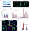
FIGURE 1.
FIGURE 1.
Effect of HES1 inhibition on redifferentiation of expanded islet cells. Cells at passage 4 were analyzed 9 days following infection of with HES1 shRNA or nontarget viruses. A, immunoblotting analyses for HES1 and p57. HSC70, heat-shock cognate 70. B, DNA staining (with DAPI) showing cell clustering. Bar = 100 μm. C, change in nucleus size. Values are mean ± S.D. (n = 1000 cells). Asterisks mark significant changes (p < 0.01). D, qPCR analyses. Relative quantification (RQ) values compared with nontarget virus are shown as mean ± S.E. (n = 3–4 donors). Asterisks mark significant changes (p < 0.01). E, HES1 inhibition induces redifferentiation of BCD cells. Immunofluorescence analysis of BCD (eGFP+) cells. Nontarget-infected cells showed no C-peptide staining. Bar = 100 μm; inset bar = 20 μm. F, qPCR analyses of RNA extracted from expanded human islet cells at passage 5, 7 days following infection with HES1 shRNA lentivirus, nontarget lentivirus, or p57 adenovirus. Values represent mean ± S.E. (n = 4 donors).
FIGURE 2.
FIGURE 2.
Effect of HES1 shRNA and SFM on morphology of BCD cells. Phase contrast and GFP fluorescence images of sorted eGFP+ and eGFP cells at passage 7 on day 8 following infection with HES1 shRNA or nontarget virus and day 4 after seeding in SFM. Bar = 100 μm.
FIGURE 3.
FIGURE 3.
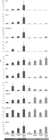
Effect of HES1 shRNA and SFM on redifferentiation of BCD cells. qPCR analysis of RNA from sorted cells infected at passage 7 with HES1 shRNA or nontarget viruses. Four days following infection, cells were seeded in SFM or growth medium. RNA was extracted 4 days later and analyzed with primers for the indicated genes. RQ values are relative to cells infected with nontarget virus and incubated in growth medium and represent mean ± S.E. (n = 4 donors). Asterisks mark significant changes (p < 0.05) induced by HES1 shRNA and SFM compared with nontarget virus-infected eGFP+ cells in growth medium.
FIGURE 4.
FIGURE 4.
Effect of HES1 shRNA and RM on redifferentiation of expanded islet cells and BCD cells. A, qPCR analysis of RNA from sorted eGFP+ cells infected at passage 5 with HES1 shRNA or nontarget viruses. Two to 4 days following infection, cells were seeded in RM. RNA was extracted 6–11 days later and analyzed with primers for the indicated genes. RQ values are relative to untreated (UTR) cells in growth medium (10% FCS) and represent mean ± S.E. (n = 4 donors). Asterisks mark significant changes (p < 0.05). B and C, co-immunofluorescence analysis for C-peptide and the indicated antigen in islet cells infected at passage 5 with HES1 shRNA and seeded 2 days later in RM for 4 days. Bar = 10 μm. B, C-peptide/eGFP co-staining in labeled cells. C, C-peptide/islet hormone co-staining in unlabeled cells. D, quantitation of 2000 cells/group in the experiment shown in C. Values are mean ± S.E. (n = 3 donors). No hormone-positive cells were found in untreated cells. Asterisks mark significant changes (p < 0.05). E, co-immunofluorescence analysis for PDX1 or NKX2.2 and C-peptide in islet cells infected at passage 5 with HES1 shRNA virus. Cells were seeded in RM 2 days following infection and stained 4 days later. Bar = 10 μm (top) and 20 μm (bottom).
FIGURE 5.
FIGURE 5.
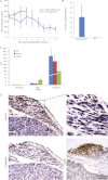
Transplantation of cells treated with HES1 shRNA into hyperglycemic immunodeficient mice corrects glycemia. NOD-SCID mice were made diabetic by streptozotocin treatment and transplanted with 2 × 10⁶ expanded islet cells infected at passage 5 with HES1 shRNA virus. A, changes in blood glucose levels in mice transplanted with cells infected with HES1 shRNA or nontarget virus. Values are mean ± S.E. B, serum human C-peptide levels 40 days following transplantation. Values are mean ± S.D. (n = 3 mice). C, transcript levels in RNA extracted from the transplants 56 days following transplantation in comparison with shRNA-infected cells in vitro. RQ values are relative to nontarget-infected cells in vitro and represent mean ± S.E. (n = 3 transplants). D, immunohistochemical analysis of transplant sections 56 days following transplantation. Dashed red lines mark transplant-kidney boundary. Bar = 100 μm.
FIGURE 6.
FIGURE 6.
Proposed model for β-cell dedifferentiation, replication, and redifferentiation. During the initial days in culture, insulin expression declines, whereas HES1 expression is induced. HES1 induction blocks p57 expression, induces β-cell replication, and causes further dedifferentiation. HES1 shRNA induces BCD cell redifferentiation following expansion. Redifferentiated cells are capable of further maturation in vivo into functional beta-like cells.

References

    1. Meier J. J., Bhushan A., Butler A. E., Rizza R. A., Butler P. C. (2005) Sustained β cell apoptosis in patients with long-standing type 1 diabetes: indirect evidence for islet regeneration? Diabetologia 48, 2221–2228 - PubMed
    1. Meier J. J., Butler A. E., Saisho Y., Monchamp T., Galasso R., Bhushan A., Rizza R. A., Butler P. C. (2008) β-cell replication is the primary mechanism subserving the postnatal expansion of β-cell mass in humans. Diabetes 57, 1584–1594 - PMC - PubMed
    1. Butler A. E., Janson J., Bonner-Weir S., Ritzel R., Rizza R. A., Butler P. C. (2003) β-Cell deficit and increased β-cell apoptosis in humans with type 2 diabetes. Diabetes 52, 102–110 - PubMed
    1. Nielsen J. H., Brunstedt J., Andersson A., Frimodt-Møller C. (1979) Preservation of β cell function in adult human pancreatic islets for several months in vitro. Diabetologia 16, 97–100 - PubMed
    1. Hayek A., Beattie G. M., Cirulli V., Lopez A. D., Ricordi C., Rubin J. S. (1995) Growth factor/matrix-induced proliferation of human adult β-cells. Diabetes 44, 1458–1460 - PubMed

Publication types

MeSH terms

LinkOut - more resources