. 2012 May 18;287(21):17353-17362.
doi: 10.1074/jbc.M111.338897. Epub 2012 Mar 28.

Endothelin induces rapid, dynamin-mediated budding of endothelial caveolae rich in ET-B

Affiliations

Endothelin induces rapid, dynamin-mediated budding of endothelial caveolae rich in ET-B

Phil Oh et al. J Biol Chem. .

Abstract

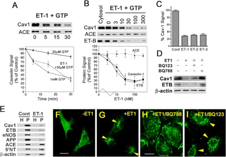
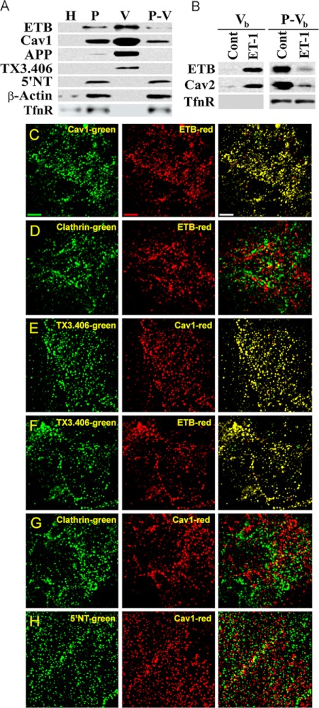
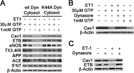
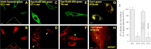
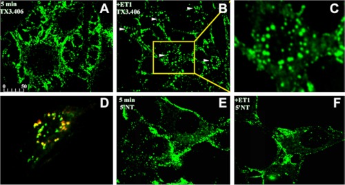
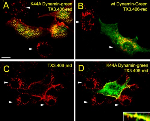
Clathrin-independent trafficking pathways for internalizing G protein-coupled receptors (GPCRs) remain undefined. Clathrin-mediated endocytosis of receptors including ligand-engaged GPCRs can be very rapid and comprehensive (<10 min). Caveolae-mediated endocytosis of ligands and antibodies has been reported to be much slower in cell culture (≫10 min). Little is known about the role of physiological ligands and specific GPCRs in regulating caveolae trafficking. Here, we find that one receptor for endothelin, ET-B but not ET-A, resides on endothelial cell surfaces in both tissue and cell culture primarily concentrated within caveolae. Reconstituted cell-free budding assays show that endothelins (ETs) induce the fission of caveolae from endothelial plasma membranes purified from rat lungs. Electron microcopy of lung tissue sections and tissue subcellular fractionation both show that endothelin administered intravascularly in rats also induces a significant loss of caveolae at the luminal surface of lung vascular endothelium. Endothelial cells in culture show that ET stimulates very rapid internalization of caveolae and cargo including caveolin, caveolae-targeting antibody, and itself. The ET-B inhibitor BQ788, but not the ET-A inhibitor BQ123, blocks the ET-induced budding of caveolae. Both the pharmacological inhibitor Dynasore and the genetic dominant negative K44A mutant of dynamin prevent this induced budding and internalization of caveolae. Also shRNA lentivirus knockdown of caveolin-1 expression prevents rapid internalization of ET and ET-B. It appears that endothelin can engage ET-B already highly concentrated in caveolae of endothelial cells to induce very rapid caveolae fission and endocytosis. This transport requires active dynamin function. Caveolae trafficking may occur more rapidly than previously documented when it is stimulated by a specific ligand to signaling receptors already located in caveolae before ligand engagement.


Figures

FIGURE 1.
FIGURE 1.
Endothelins induce endothelial caveolae budding in vitro, in cell culture, and in situ. A–D, in vitro reconstituted cell-free budding assay (see “Experimental Procedures”). Western blot analysis of repelleted silica-coated endothelial plasma membranes showing the signal for the indicated proteins either after 100 nm ET-1 stimulation for the indicated times (A) or stimulation with indicated concentration (nm) of ET-1 for 30 min (B). The graphs represent the quantified signal from the Western blot analysis as a percentage of control signal (either ET-1 at time 0 or no ET-1, respectively) for the indicated protein and then plotted as a function of incubation time (A) or ET-1 concentration (B). C, histogram showing relative cav1 signal remaining on repelleted silica-coated endothelial plasma membranes after 30 min of treatment with of ET-1, ET-2, and ET-3. D, Western blot analysis of the repelleted silica-coated endothelial plasma membrane proteins showing the signal for the indicated primary antibodies after 30 min of pretreatment with either 50 nm BQ123 (ET-A antagonist) or 5 nm BQ788 (ET-B antagonist) followed by 30 min of stimulation with ET-1. E, Western blot analysis of silica-coated endothelial plasma membranes isolated from rat lungs showing the signal for the indicated primary antibodies after 5 min of stimulation with ET-1. F–I, confocal fluorescence microscopic images of cav1-eGFP expressing BAEC showing caveolin-1 localization either untreated (F) or after 5 min of stimulation with ET-1 (G). Confocal fluorescence microscopic images of BAEC showing localization of caveolin-1 after pretreated for 30 min with either 5 nm BQ788 (H) or 50 nm BQ123 (I) followed by 5 min of stimulation with ET-1. The arrowheads indicate cav1 perinuclear intracellular localization. H, total lung homogenate; eNOS, endothelial nitric-oxide synthase. Cont, control. All of the graphs are the percentages of the control cav1 signal detected without ET1 exposure. n ≥ 3 experiments. The bar represents 100 μm.
FIGURE 2.
FIGURE 2.
ET-B is concentrated specifically in dynamic caveolae capable of budding to form free transport vesicles. A, Western blot analysis of subcellular fractions (1 μg) isolated from rat lung tissue (see “Experimental Procedures”). H, whole lung homogenates; P-V, repelleted silica-coated membranes stripped of caveolae. B, Western blot analysis of caveolae induced to bud by ET-1 in the in vitro reconstituted budding assay (see “Experimental Procedures”). P either untreated (control, Cont) or treated with ET-1 for 30 min at 37 °C was subjected to sucrose density centrifugation to separate the low density caveolae vesicles released from P (Vb) from the repelleted silica-coated endothelial plasma membrane proteins (P-Vb). C–H, dual immunofluorescence microscopy of intact RLMVEC incubated at 4 °C with antibodies (see “Experimental Procedures”) as indicated. Single channel green and red fluorescence images are shown along with the merged images with both signals. Colocalization of signals generates yellow in the overlay of the two images. Bar, 2 μm. TfnR, transferrin receptor; Cav2, caveolin-2.
FIGURE 3.
FIGURE 3.
Role of dynamin in caveolae fission induced by ET-1. A, Western blot analysis of repelleted endothelial cell plasma membranes after performing the in vitro reconstituted budding assay in the presence of ET-1 for 30 min, GTP, and either wild type (wt) or K44A mutant dynamin containing cytosol as indicated (see “Experimental Procedures”). B, same as A but used standard cytosol and as indicated with 50 μm Dynasore (30 min of pretreatment before ET-1 and also included with ET-1). C, Western analysis of silica-coated endothelial plasma membranes isolated from rat lungs that had been treated in situ (see “Experimental Procedures”) with ET-1 and/or 50 μm Dynasore. Dynasore was administered for 10 min and then also combined with ET-1 as indicated.
FIGURE 4.
FIGURE 4.
ET-1 is rapidly internalized by caveolae in cultured endothelial cells. Images from confocal fluorescence microscopy of tetramethylrhodamine-ET1 localization (red) on intact BAEC immediately fixed after 4 °C binding (A) or after the cells were warmed to 37 °C for 5 min (B) prior to fixation with paraformadehyde. The cells were then permeabilized before incubation with cav1 antibodies and fluorophore-conjugated reporter antibodies (cav1, green). The bar represents 50 μm.
FIGURE 5.
FIGURE 5.
Role of dynamin and caveolin-1 but not Eps15 in rapid internalization of ET-1 and ET-B in cultured endothelial cells. A–F, images from confocal fluorescence microscopy of intact BAEC expressing either K44A Dynamin2-eGFP (green) (A and B) or Eps15-eGFP mutant (green) (C–F), and localization of either tetramethylrhodamine-ET1 (red) (A–D) or tetramethylrhodamine-Tfn (red) (E and F) after binding at 4 °C followed by warming to 37 °C for 5 min prior to fixation with paraformaldehyde (see “Experimental Procedures”). The dashed lines in A and B outline the positions of untransfected cell. The arrowheads in D and F denote Eps15 mutant transfected cells. G and H, localization of cav1 and ETB after 5 min of stimulation with ET1 of BAEC that were uninfected (G) or already infected for 24 h with shCav1 (H). I, histogram showing relative biotinylated ET-1 cell surface signal detected using an enzyme-linked, avidin-based detection assay (see “Experimental Procedures”). Intact, nonfixed BAEC were incubated with biotinylated ET-1 at 4 °C. Some cells (labeled 4 °C) were immediately fixed and processed for biotin detection. The other cells (labeled 37 °C) were first warmed to 37 °C for 5 min before processing. The cells infected for 24 h with shRNA lentivirus transduction particle are indicated as either +shCav1 for caveolin-1 shRNA or with control shRNA lentivirus transduction particle (+shCont) for control shRNA. *, p < 0.05 (analysis of variance/Tukey's range test). n ≥ 3 experiments of triplicate wells for each experiment. The bar represents 50 μm.
FIGURE 6.
FIGURE 6.
ET-1-induced internalization of TX3.406, an antibody targeting caveolae, in cultured cells. Images from confocal fluorescence microscopy of intact RLMVEC incubated at 4 °C with either TX3.406 (A–D) or 5′NT antibody (E and F) to label caveolae and lipid rafts, respectively. The cells were then incubated for 5 min at 37 °C in the presence (B–D and F) or absence (A and E) of ET-1 and then fixed and processed with fluorescent reporter antibody (green). C, magnified image of cell indicated by arrow and yellow box in B. D, the RLMVEC stimulated with ET-1 were also permeabilized and incubated with polyclonal antibody to caveolin-1 followed by fluorescent reporter antibody to rabbit IgG (red). The arrowheads indicate internalized signal. The bar represents 50 μm (A, B, and D–F) or 10 μm (C).
FIGURE 7.
FIGURE 7.
Role of dynamin in ET-1 induced internalization of TX3.406 in cultured endothelial cells. Images from confocal fluorescence microscopy of RLMVEC already transfected to express either K44A dynamin2-eGFP (green) (A, C, and D) or wild type (wt) dynamin2-eGFP (green) (B). The cells were incubated with TX3.406 for 1 h at 4 °C and washed before 5 min of stimulation with ET-1 at 37 °C and then fixation, permeabilization, and incubation with secondary fluorescent reporter antibody to mouse IgG (red). The arrowheads indicated intracellular perinuclear staining. Bar, 10 μm.

References

    1. Montesano R., Roth J., Robert A., Orci L. (1982) Non-coated membrane invaginations are involved in binding and internalization of cholera and tetanus toxins. Nature 296, 651–653 - PubMed
    1. Parton R. G., Joggerst B., Simons K. (1994) Regulated internalization of caveolae. J. Cell Biol. 127, 1199–1215 - PMC - PubMed
    1. Schnitzer J. E., Oh P., McIntosh D. P. (1996) Role of GTP hydrolysis in fission of caveolae directly from plasma membranes. Science 274, 239–242 - PubMed
    1. Pelkmans L., Kartenbeck J., Helenius A. (2001) Caveolar endocytosis of simian virus 40 reveals a new two-step vesicular-transport pathway to the ER. Nat. Cell Biol. 3, 473–483 - PubMed
    1. Schnitzer J. E., Oh P., Pinney E., Allard J. (1994) Filipin-sensitive caveolae-mediated transport in endothelium: reduced transcytosis, scavenger endocytosis, and capillary permeability of select macromolecules. J. Cell Biol. 127, 1217–1232 - PMC - PubMed

Publication types

MeSH terms