. 2012 Mar 28;32(13):4334-40.
doi: 10.1523/JNEUROSCI.5845-11.2012.

Bidirectional relationship between functional connectivity and amyloid-β deposition in mouse brain

Affiliations

Bidirectional relationship between functional connectivity and amyloid-β deposition in mouse brain

Adam W Bero et al. J Neurosci. .

Abstract

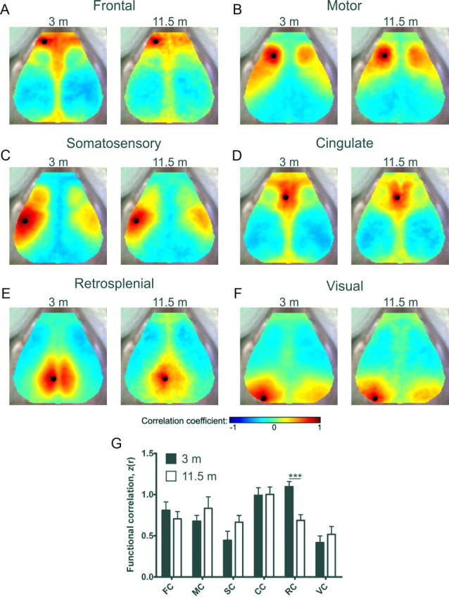
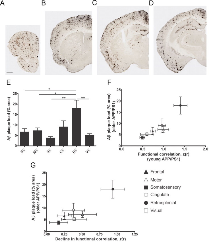
Brain region-specific deposition of extracellular amyloid plaques principally composed of aggregated amyloid-β (Aβ) peptide is a pathological signature of Alzheimer's disease (AD). Recent human neuroimaging data suggest that resting-state functional connectivity strength is reduced in patients with AD, cognitively normal elderly harboring elevated amyloid burden, and in advanced aging. Interestingly, there exists a striking spatial correlation between functional connectivity strength in cognitively normal adults and the location of Aβ plaque deposition in AD. However, technical limitations have heretofore precluded examination of the relationship between functional connectivity, Aβ deposition, and normal aging in mouse models. Using a novel functional connectivity optical intrinsic signal (fcOIS) imaging technique, we demonstrate that Aβ deposition is associated with significantly reduced bilateral functional connectivity in multiple brain regions of older APP/PS1 transgenic mice. The amount of Aβ deposition in each brain region was associated with the degree of local, age-related bilateral functional connectivity decline. Normal aging was associated with reduced bilateral functional connectivity specifically in retrosplenial cortex. Furthermore, we found that the magnitude of regional bilateral functional correlation in young APP/PS1 mice before Aβ plaque formation was proportional to the amount of region-specific plaque deposition seen later in older APP/PS1 mice. Together, these findings suggest that Aβ deposition and normal aging are associated with region-specific disruption of functional connectivity and that the magnitude of local bilateral functional connectivity predicts regional vulnerability to subsequent Aβ deposition in mouse brain.


Figures

Figure 1.
Figure 1.
Aβ plaque deposition is associated with decreased functional connectivity in multiple brain systems of older APP/PS1 mice. A–F, Composite, group-averaged, functional correlation maps of frontal (FC, A), motor (MC, B), somatosensory (SC, C), cingulate (CC, D), retrosplenial (RC, E) and visual (VC, F) cortices in young and older APP/PS1 mice. Black circles denote seed position. G, Regional bilateral functional correlation in young (black bars) and older (white bars) APP/PS1 mice (n = 7/group). *p < 0.05, ***p < 0.001. Values represent mean ± SEM. H, Locations of cortical functional regions, anatomical landmarks (white circles), and seed placement and size (black circles) were manually constructed (White et al., 2011) using the Franklin and Paxinos (1996) histological atlas.
Figure 2.
Figure 2.
Normal aging is associated with decreased functional connectivity in retrosplenial cortex. A–F, Composite, group-averaged, functional correlation maps of frontal (FC, A), motor (MC, B), somatosensory (SC, C), cingulate (CC, D), retrosplenial (RC, E) and visual (VC, F) cortices in young and older wild-type mice. Black circles denote seed position. G, Regional bilateral functional correlation in young (black bars) and older (white bars) wild-type mice (young, n = 10; older, n = 13); ***p < 0.001. Values represent mean ± SEM.
Figure 3.
Figure 3.
Regional bilateral functional connectivity in APP/PS1 mice is predictive of and affected by regional plaque deposition. A–D, Representative brain sections from older APP/PS1 mice stained with biotinylated HJ3.4 antibody (anti-Aβ1–13) to visualize Aβ immunopositive plaques (n = 4/group). Scale bar: (in A) AD, 500 μm. E, Percentage area occupied by Aβ deposition in frontal (FC), motor (MC), somatosensory (SC), cingulate (CC), retrosplenial (RC), and visual (VC) cortices; *p < 0.05, **p < 0.01. F, Regional bilateral correlation in young APP/PS1 mice plotted against Aβ plaque deposition in each brain region of older APP/PS1 mice. G, Decline in bilateral correlation of older APP/PS1 mice plotted against Aβ plaque deposition. Values represent mean ± SEM.
Figure 4.
Figure 4.
Aβ deposition is associated with degree of decline in local bilateral functional connectivity. A, B, Consensus bilateral functional connectivity maps generated for young (A) and older (B) APP/PS1 mice and young (E) and older (F) wild-type mice. C, G, Bilateral functional connectivity difference map between young and older APP/PS1 (C) and wild-type (G) mice. D, H, p values of C, G, plotted on a −log10 scale. Note that in C, G, positive values represent age-related decline, whereas negative values represent age-related increase in bilateral functional connectivity. White circles denote anatomical landmarks for image coregistration.

References

    1. Andrews-Hanna JR, Snyder AZ, Vincent JL, Lustig C, Head D, Raichle ME, Buckner RL. Disruption of large-scale brain systems in advanced aging. Neuron. 2007;56:924–935. - PMC - PubMed
    1. Bero AW, Yan P, Roh JH, Cirrito JR, Stewart FR, Raichle ME, Lee JM, Holtzman DM. Neuronal activity regulates the regional vulnerability to amyloid-beta deposition. Nat Neurosci. 2011;14:750–756. - PMC - PubMed
    1. Buckner RL, Snyder AZ, Shannon BJ, LaRossa G, Sachs R, Fotenos AF, Sheline YI, Klunk WE, Mathis CA, Morris JC, Mintun MA. Molecular, structural, and functional characterization of Alzheimer's disease: evidence for a relationship between default activity, amyloid, and memory. J Neurosci. 2005;25:7709–7717. - PMC - PubMed
    1. Buckner RL, Sepulcre J, Talukdar T, Krienen FM, Liu H, Hedden T, Andrews-Hanna JR, Sperling RA, Johnson KA. Cortical hubs revealed by intrinsic functional connectivity: mapping, assessment of stability, and relation to Alzheimer's disease. J Neurosci. 2009;29:1860–1873. - PMC - PubMed
    1. Corcoran KA, Donnan MD, Tronson NC, Guzmán YF, Gao C, Jovasevic V, Guedea AL, Radulovic J. NMDA receptors in retrosplenial cortex are necessary for retrieval of recent and remote context fear memory. J Neurosci. 2011;31:11655–11659. - PMC - PubMed

Publication types

Substances

LinkOut - more resources