. 2012 Mar 28;32(13):4675-87.
doi: 10.1523/JNEUROSCI.4749-11.2012.

Photoreceptor coupling mediated by connexin36 in the primate retina

Affiliations

Photoreceptor coupling mediated by connexin36 in the primate retina

Jennifer J O'Brien et al. J Neurosci. .

Abstract

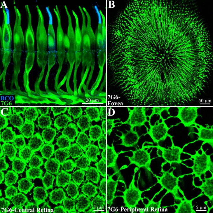
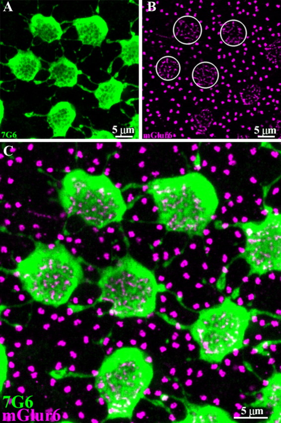
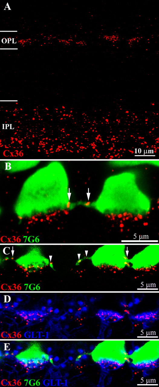
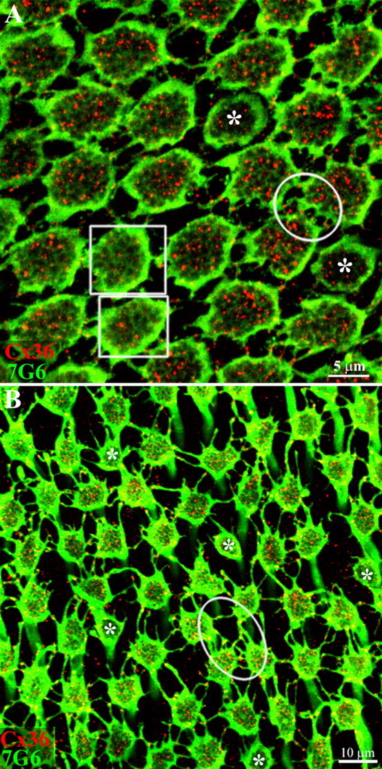
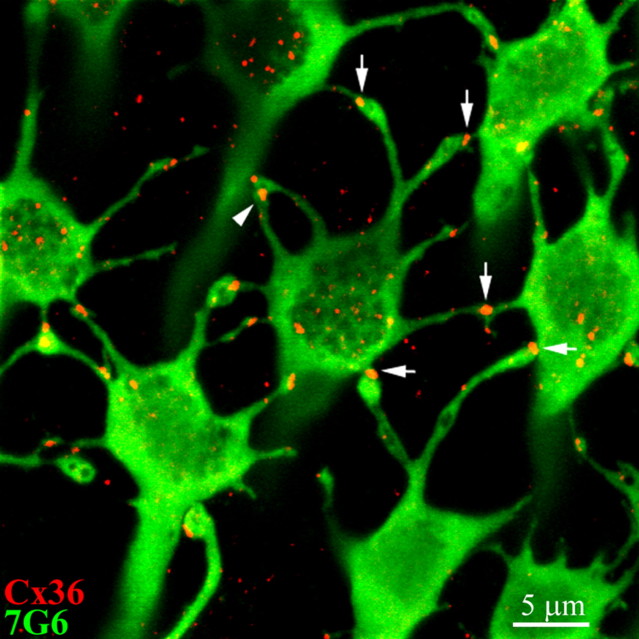
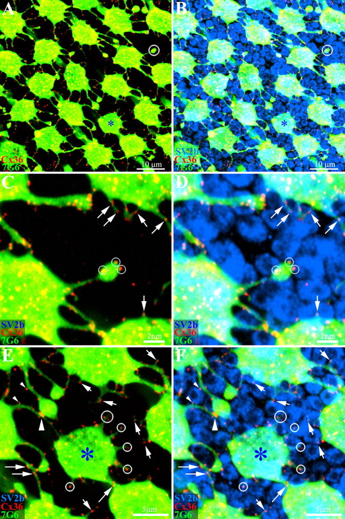
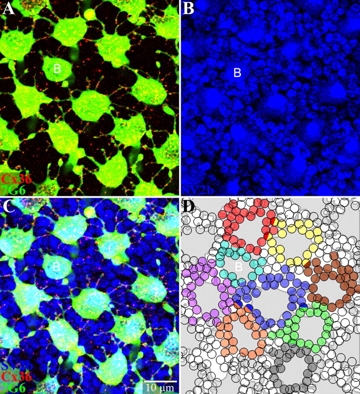
Photoreceptors are coupled via gap junctions in many mammalian species. Cone-to-cone coupling is thought to improve sensitivity and signal-to-noise ratio, while rod-to-cone coupling provides an alternative rod pathway active under twilight or mesopic conditions (Smith et al., 1986; DeVries et al., 2002; Hornstein et al., 2005). Gap junctions are composed of connexins, and connexin36 (Cx36), the dominant neuronal connexin, is expressed in the outer plexiform layer. Primate (Macaca mulatta) cone pedicles, labeled with an antibody against cone arrestin (7G6) were connected by a network of fine processes called telodendria and, in double-labeled material, Cx36 plaques were located precisely at telodendrial contacts between cones, suggesting strongly they are Cx36 gap junctions. Each red/green cone made nonselective connections with neighboring red/green cones. In contrast, blue cone pedicles were smaller with relatively few short telodendria and they made only rare or equivocal Cx36 contacts with adjacent cones. There were also many smaller Cx36 plaques around the periphery of every cone pedicle and along a series of very fine telodendria that were too short to reach adjacent members of the cone pedicle mosaic. These small Cx36 plaques were closely aligned with nearly every rod spherule and may identify sites of rod-to-cone coupling, even though the identity of the rod connexin has not been established. We conclude that the matrix of cone telodendria is the substrate for photoreceptor coupling. Red/green cones were coupled indiscriminately but blue cones were rarely connected with other cones. All cone types, including blue cones, made gap junctions with surrounding rod spherules.


Figures

Figure 1.
Figure 1.
An antibody to cone arrestin, 7G6, labels primate cones. A, Cones in the macaque retinal sections were labeled with 7G6, a cone arrestin antibody, shown in green. Cones were labeled in their entirety with particularly bright staining of the outer segments and pedicles or synaptic terminals. Red/green cones cannot be distinguished but double labeling with an antibody against blue cone opsin (blue) shows that blue cones are also stained by 7G6. Optical sections 8 × 0.5 μm. B, A whole-mount view of the macaque fovea shows cones labeled with the 7G6 antibody. The axons of the foveal cones are displaced radially away from the foveal pit, forming Henle's fiber layer, and terminating in a ring of cone pedicles around the fovea. A 20× objective, 9 × 1 μm sections. C, Central primate retina, focus at the level of the outer plexiform layer. Cone pedicles, labeled with 7G6, are densely packed, often in contact. They also extend short processes, called telodendria, to contact adjacent pedicles. The rim of each cone pedicle is densely stained but there are many holes in the base of the cone pedicle that correspond to synaptic invaginations (see below). Optical sections 5 × 0.4 μm. D, In peripheral primate retina, the cone density decreases, the cone pedicles are widely spaced and the telodendria lengthen to compensate. Optical sections 5 × 0.4 μm.
Figure 2.
Figure 2.
7G6 antibodies outline synaptic invagination sites. A, A high-resolution image of 7G6-labeled cones shows several cone pedicles interconnected by telodendria. Some cone telodendria appear to have enlarged endings in peripheral retina. As in Figure 1, there are holes in the central base of each cone pedicle. B, The tips of ON cone bipolar cells and rod bipolar cells contain mGluR6. Antibodies against mGluR6 (magenta) stain two distinct structures in the outer plexiform layer. The tips of ON cone bipolar cells appear as clusters of small mGluR6-labeled processes, which invaginate the cone pedicle (several circled). The tips of rod bipolar cells appear as brightly stained doublets that occupy the space between cone pedicles. These also indicate the positions of unlabeled rod spherules. C, Double-label image shows that the holes in the center of 7G6-labeled cone pedicles colocalize with the mGluR6 signals of ON cone bipolar cells. This indicates that the dendritic endings of ON cone bipolar cells enter each hole in the base of the cone pedicle. Therefore, the holes represent synaptic invagination sites and indicate that the level of focus is at the very base of the cone pedicle. Optical sections 5 × 0.5 μm.
Figure 3.
Figure 3.
Cx36 labeling occurs both between and underneath cone pedicles. A, A cross section of macaque retina shows Cx36 labeling in both plexiform layers. The prominent Cx36 labeling in the inner IPL is mainly associated with AII amacrine cells. In comparison, the Cx36 labeling in the OPL is fainter; the plaques are smaller and form clusters. B, Double labeling shows that Cx36 plaques occur in two locations: on fine processes between 7G6-labeled cone pedicles (arrows) and in prominent clusters underneath each cone pedicle. The Cx36 plaques underneath the cone pedicle are distinct from and do not colocalize with the base of the cone pedicle. C–E, Triple label images from vertical sections of macaque retina. C, Cone pedicles are stained with 7G6 (green). Cx36 plaques (red) occur at cone pedicle contacts (arrows) and on fine processes leaving the cone pedicle base (arrowheads). D, Certain OFF bipolar cells, including OFF midget bipolar cells and DB2, are labeled with antibodies against the glutamate transporter GLT-1 (blue). The OFF bipolar dendrites terminate in flat tops, apposed to the base of the cone pedicle. Most of the Cx36 labeling underneath the cone pedicles is colocalized with the OFF bipolar dendrites. E, Triple label image shows that the Cx36 plaques below each cone pedicle are colocalized with GLT-1 stained OFF bipolar cells and not with the cone pedicle. All panels, 6 × 0.4 μm optical sections.
Figure 4.
Figure 4.
Cx36 forms gap junctions between red/green cones. Macaque retina was viewed in whole-mount with the level of focus at the base of the cone pedicles in the OPL. A, In central retina, cone pedicles stained for 7G6 (green) were tightly packed, frequently with direct contacts between neighboring pedicles. In addition, there are many small processes or telodendria, which contact adjacent pedicles. The Cx36 labeling within the perimeter of each cone pedicle is due to the presence of Cx36 gap junctions beneath the cone pedicle (red). In two pedicles (+) (boxes) that are slightly higher, the synaptic invaginations (holes) in the pedicle base begin to blur and fill in and the Cx36 labeling underneath is markedly reduced. Two smaller, potentially blue cone pedicles are marked with asterisks. It should be noted that Cx36 plaques occur at nearly all direct or telodendrial contacts between red/green cone pedicles. For example, a well labeled group of Cx36 gap junctions are stained within the circle. Optical sections 6 × 0.4 μm. B, In peripheral retina, the cone pedicles are more widely spaced and the telodendria are more obvious. Again, Cx36 plaques occur at telodendrial contact points between neighboring cone pedicles (oval). A few presumed blue cone pedicles, marked with asterisks, have very few telodendria or Cx36 contacts. Optical sections 6 × 0.4 μm.
Figure 5.
Figure 5.
Cx36 plaques occur precisely at telodendrial contacts between adjacent cone pedicles. A high-magnification image of 7G6-labeled cones (green) shows Cx36 (red) forms gap junctions at all 10 cone contacts of a single cone pedicle. This cone has Cx36 plaques at 8 telodendrial contacts (vertical arrows) and at 2 basal contacts from neighboring cones. The number of gap junctions may exceed the number of telodendria because some neighboring telodendria make gap junctions with the base of the cone pedicle (horizontal arrow). In addition, autologous telodendrial contacts may also occur where one telodendria forms a gap junction with another telodendria, originating from the same cone pedicle (arrowhead). Some telodendria on the left side make contacts outside the image plane. Optical sections 6 × 0.31 μm.
Figure 6.
Figure 6.
Blue cones form smaller and fewer Cx36 gap junctions. A, The outer segments of primate cones were labeled with a cone arrestin antibody (7G6). In addition, blue cone outer segments were stained with antibodies against blue cone opsin (BCO). B, The labeling could be followed through the confocal stack down to the cone cell bodies. C, The blue cone opsin and 7G6 labeling were followed further in the confocal series to identify blue cone pedicles in the array of green/red cone pedicles. Blue cone pedicles, marked by asterisks, were smaller with fewer telodendria. There was robust Cx36 labeling (red) at telodendrial contacts between cones, some of which are circled. In contrast, the telodendria of blue cones were mostly too short to reach adjacent cones. Two longer telodendria were not labeled for Cx36 (arrows).
Figure 7.
Figure 7.
Primate cones make Cx36 gap junctions with rod spherules. Whole-mount macaque retina was stained with antibodies against cone arrestin (7G6, green), Cx36 (red) and SV2b (blue), to stain rod spherules. A, A mosaic of cone pedicles in peripheral retina. A blue cone pedicle is marked with an asterisk. As before, there are many Cx36 plaques at telodendrial contacts. In addition, a few telodendria terminate in isolation (circle, top right). B, The same field shows that the space between cone pedicles is mostly filled by rod spherules stained for SV2b. C, High-magnification image of the isolated telodendron from A shows clear Cx36 plaques, even in the absence of adjoining cones pedicles (small circles). In addition, many other small telodendria, also bearing Cx36 plaques, project from a cone pedicle base (arrows). D, The isolated cone telodendron is surrounded by rod spherules and the Cx36 plaques occur at contact points with rod spherules (small circles). The Cx36 plaques on short telodendria also contact rod spherules (arrows). E, The blue cone pedicle also has many fine telodendria, too short to reach adjacent cones. One that approaches a nearby cone has Cx36 plaques aligned with rod spherules (diagonal arrow). In addition, Cx36 plaques may be observed along a telodendrial shaft (long arrows). F, Cx36 plaques on the blue cone telodendria are aligned with the surrounding rod spherules (circles). Some rod spherules have Cx36 contacts with the blue cone pedicle and an adjacent cone pedicle. Two small Cx36 plaques from different telodendria contact opposite sides of a single rod spherule (small arrowheads). Further down, there is a much larger cone-to-cone gap junction (large arrowhead). In summary, many fine telodendria form all types of cones make small Cx36 gap junctions with surrounding rod spherules (small arrows). A–D, 4 × 0.4 μm optical sections; E, F, 3 × 0.4 μm optical sections.
Figure 8.
Figure 8.
Cone pedicles make potential Cx36 gap junctions with most surrounding rod spherules. A, A mosaic of cone pedicles stained with 7G6 (green). Many telodendria, long and short, are decorated with Cx36 plaques (red). A single small blue cone pedicle is marked with a letter B. B, Same field showing rod spherules, which are round with a dark central spot stained for SV2b (blue). Cone pedicles are larger polygonal structures. C, Triple-label image shows Cx36 contacts with many neighboring rods and cones. D, A reconstruction of part of the cone pedicle mosaic. The cone pedicles were outlined and colored gray. Each rod spherule receiving a Cx36 telodendrial contact was shaded with one color indicating contacts from a single cone pedicle. A few rods receiving Cx36 contacts from two different cones were coded as hemispheres of two colors, including at least two rod spherules with blue input. The small telodendria extend for 2–3 rows of rod spherules around each cone pedicle. Almost every rod spherule receives potential gap junction contacts. Red/green cone pedicles contacted 20–30 adjacent rods. A single blue cone pedicle (B) made potential contacts with 16 rod spherules. Optical sections 6 × 0.3 μm.

Similar articles

Cited by

References

    1. Abd-El-Barr MM, Pennesi ME, Saszik SM, Barrow AJ, Lem J, Bramblett DE, Paul DL, Frishman LJ, Wu SM. Genetic dissection of rod and cone pathways in the dark-adapted mouse retina. J Neurophysiol. 2009;102:1945–1955. - PMC - PubMed
    1. Ahnelt PK, Kolb H. The mammalian photoreceptor mosaic-adaptive design. Prog Retin Eye Res. 2000;19:711–777. - PubMed
    1. Ahnelt PK, Kolb H, Pflug R. Identification of a subtype of cone photoreceptor, likely to be blue sensitive, in the human retina. J Comp Neurol. 1987;255:18–34. - PubMed
    1. Ahnelt P, Keri C, Kolb H. Identification of pedicles of putative blue-sensitive cones in the human retina. J Comp Neurol. 1990;293:39–53. - PubMed
    1. Attwell D, Borges S, Wu SM, Wilson M. Signal clipping by the rod output synapse. Nature. 1987;328:522–524. - PubMed

Publication types