. 2012 Jun;86(11):6315-22.
doi: 10.1128/JVI.00050-12. Epub 2012 Mar 28.

Hepatitis C virus activates the mTOR/S6K1 signaling pathway in inhibiting IRS-1 function for insulin resistance

Affiliations

Hepatitis C virus activates the mTOR/S6K1 signaling pathway in inhibiting IRS-1 function for insulin resistance

Sandip K Bose et al. J Virol. 2012 Jun.

Abstract

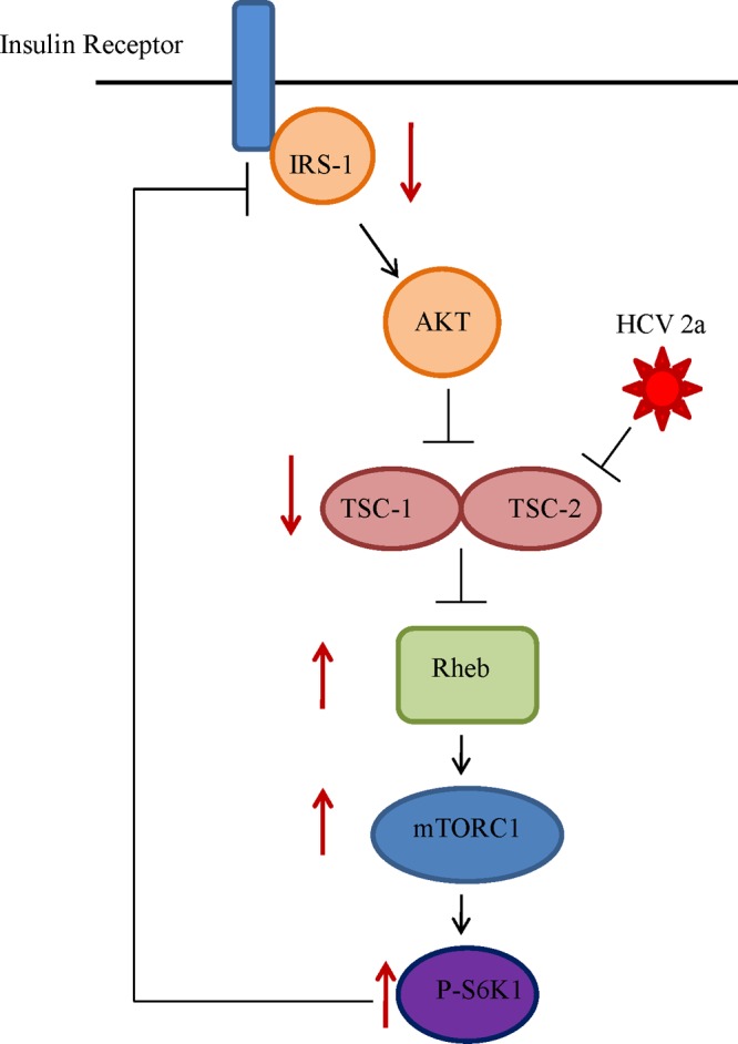
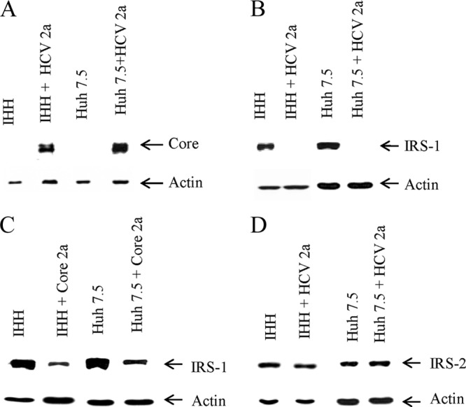
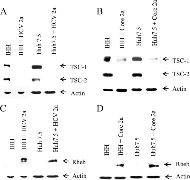
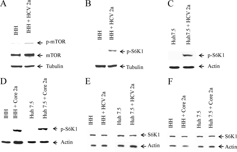
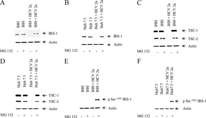
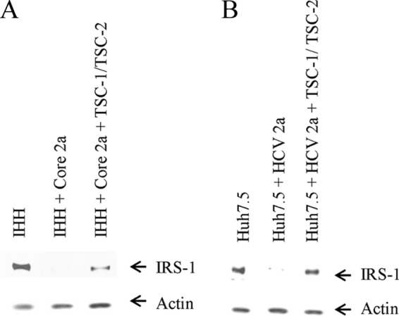
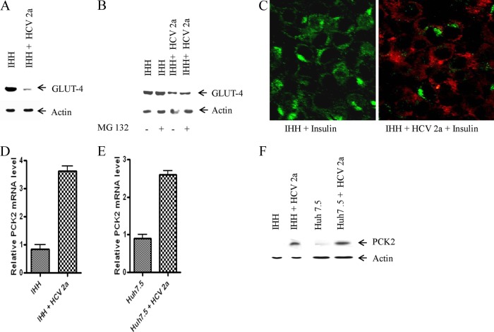
Hepatitis C virus (HCV) infection significantly increases the prevalence of type 2 diabetes mellitus (T2DM). Insulin receptor substrate 1 (IRS-1) plays a key role in insulin signaling, thus enabling metabolic regulation in mammalian cells. We have previously shown that HCV infection modulates phosphorylation of Akt, a downstream target of IRS-1. In this study, we further examined the status of total IRS-1 and the downstream regulation of the Akt pathway in understanding mTOR/S6K1 signaling using HCV genotype 2a (clone JFH1)-infected hepatocytes. Inhibition of IRS-1 expression was observed in HCV-infected hepatocytes compared to that in a mock-infected control. The status of the tuberous sclerosis complex (TSC-1/TSC-2) was significantly decreased after HCV infection of human hepatocytes, showing a modulation of the downstream Akt pathway. Subsequent study indicated an increased level of Rheb and mTOR expression in HCV-infected hepatocytes. Interestingly, the phosphoS6K1 level was higher in HCV-infected hepatocytes, suggesting a novel mechanism for IRS-1 inhibition. Ectopic expression of TSC-1/TSC-2 significantly recovered the IRS-1 protein expression level in HCV-infected hepatocytes. Further analyses indicated that HCV core protein plays a significant role in modulating the mTOR/S6K1 signaling pathway. Proteasome inhibitor MG 132 recovered IRS-1 and TSC1/2 expression, suggesting that degradation occurred via the ubiquitin proteasome pathway. A functional consequence of IRS-1 inhibition was reflected in a decrease in GLUT4 protein expression and upregulation of the gluconeogenic enzyme PCK2 in HCV-infected hepatocytes. Together, these observations suggested that HCV infection activates the mTOR/S6K1 pathway in inhibiting IRS-1 function and perturbs glucose metabolism via downregulation of GLUT4 and upregulation of PCK2 for insulin resistance.


Figures

Fig 1
Fig 1
Simplified schematic presentation of HCV genotype 2a-mediated blocking of the insulin signaling pathway. We suggested previously that HCV infection upregulates Ser-312 phosphorylation of IRS-1 and impairs the Akt signaling pathway by modulation of Ser/Thr phosphorylation in the upstream insulin signaling pathway (11). Analysis of the downstream signaling pathway reveals that HCV further downregulates tumor suppressor TSC1/TSC2, resulting in an upregulation of Rheb. This results in downstream activation of mTOR and S6K1, which in turn acts as a negative regulator of IRS-1, causing insulin resistance. Directions of solid arrows on the side indicate up- or downregulation of specific signaling molecules.
Fig 2
Fig 2
IRS-1 expression is markedly reduced upon HCV infection or HCV core protein expression. (A) Expression status of HCV core protein in HCV genotype 2a-infected IHH or Huh7.5 cells is shown. (B) IRS-1 status in cell culture-grown HCV genotype 2a-infected IHH or Huh7.5 cells. The expression status of actin in each lane was determined as a loading control. Mock-infected cells were included as a negative control. (C) IRS-1 protein expression status in IHH and Huh7.5 cells following transient transfection of HCV core. (D) IRS-2 expression status in HCV genotype 2a-infected IHH and Huh7.5 cells.
Fig 3
Fig 3
HCV genotype 2a infection or introduction of core gene inversely correlates with the TSC1/TSC2 and Rheb expression status. (A) Mock- or HCV genotype 2a-infected IHH or Huh7.5 cells were analyzed for TSC-1/TSC-2 expression status by Western blot analysis. The expression level of actin in each lane was determined as a loading control. (B) The TSC-1/TSC-2 expression status from HCV core 2a-transfected IHH and Huh7.5 cells is shown. (C) IHH and Huh7.5 cells infected with HCV genotype 2a were similarly analyzed for the expression level of Rheb. The expression status of actin in each lane was used as a loading control. (D) The expression status of Rheb from HCV core 2a-transfected IHH or Huh7.5 cells is shown.
Fig 4
Fig 4
HCV genotype 2a infection enhances expression of mTOR and p-S6K1. (A) Mock- and HCV genotype 2a-infected IHH cells were analyzed for total and Ser-2448 phosphorylation status of mTOR. The tubulin level in each lane was determined as a loading control. (B and C) Activated phosphorylated-S6K1 (Thr-389) status in mock- or HCV genotype 2a-infected IHH or Huh7.5 cells, respectively, were analyzed by Western blot. The tubulin or actin level in each lane was used for comparison of protein loads. (D) p-S6K1 expression status from HCV core 2a-transfected IHH or Huh7.5 cells is shown. (E) Total S6K1 status in mock-infected or HCV genotype 2a-infected IHH or Huh7.5 cells were analyzed by Western blotting. (F) Total S6K1 expression status from HCV core 2a-transfected IHH or Huh7.5 cells is also shown.
Fig 5
Fig 5
IRS-1 and TSC-1/TSC-2 are proteasomally degraded via the ubiquitin pathway. (A and B) IRS-1 status was determined in IHH and Huh7.5 cells infected with HCV genotype 2a in the presence or absence of proteasome inhibitor MG 132 (10 μM). Mock-infected cells were similarly treated with or without MG132. (C and D) TSC-1/TSC-2 status was determined in IHH and Huh7.5 cells infected with HCV genotype 2a in the presence or absence of proteasome inhibitor MG 132. Mock-infected cells were similarly treated with or without MG132. (E and F) IRS-1 Ser-1101 phosphorylation by HCV genotype 2a. Phosphorylation status of IRS-1 Ser-1101 in mock- or HCV 2a-infected IHH or Huh7.5 cells with or without MG132 treatment. The actin level was determined as a loading control.
Fig 6
Fig 6
Ectopic expression of TSC-1/TSC-2-rescued IRS-1 protein expression. (A and B) Western blot analyses displaying increased IRS-1 protein levels upon ectopic expression of TSC-1/TSC-2 in IHH transfected with HCV core 2a or Huh7.5 cells infected with HCV genotype 2a. The actin level was determined as a loading control.
Fig 7
Fig 7
HCV genotype 2a infection represses GLUT-4 expression and enhances gluconeogenic enzyme PCK2 mRNA expression level. (A) Mock- and HCV genotype 2a-infected IHH cells were analyzed for GLUT-4 protein expression by Western blotting. The actin levels were determined in each lane as a loading control. (B) Mock and HCV genotype 2a-infected IHH were analyzed for GLUT-4 protein expression by Western blot in the presence and absence of MG132 (10 μM). Actin levels were determined in each lane as a loading control. (C) Immunofluorescence displaying cellular localization of GLUT-4 in HCV genotype 2a-infected IHH. Mock- or HCV-infected cells were treated with insulin (200 nM) after 72 h of infection. Green indicates localization of GLUT4, and red indicates HCV core protein expression. (D) Relative PCK2 mRNA expression status in mock- and HCV genotype 2a-infected IHH cells. (E) A similar analysis for PCK2 mRNA expression from Huh7.5 cells is shown. The results were normalized to endogenous 18S rRNA. (F) The protein level of PCK2 was determined by Western blotting in IHH and Huh7.5 cells infected with HCV genotype 2a.

References

    1. Adinolfi LE, Durante-Mangoni E, Zampino R, Ruggiero G. 2005. Review article: hepatitis C virus-associated steatosis—pathogenic mechanisms and clinical implications. Aliment. Pharmacol. Ther. 2(Suppl):52–55 - PubMed
    1. Adinolfi LE, et al. 2001. Steatosis accelerates the progression of liver damage of chronic hepatitis C patients and correlates with specific HCV genotype and visceral obesity. Hepatology 33:1358–1364 - PubMed
    1. Aguirre V, et al. 2002. Phosphorylation of Ser307 in insulin receptor substrate-1 blocks interactions with the insulin receptor and inhibits insulin action. J. Biol. Chem. 277:1531–1537 - PubMed
    1. Aguirre V, Uchida T, Yenush L, Davis R, White MF. 2000. The c-Jun NH2-terminal kinase promotes insulin resistance during association with insulin receptor substrate-1 and phosphorylation of Ser(307). J. Biol. Chem. 275:9047–9054 - PubMed
    1. Alessi DR, et al. 1996. Mechanism of activation of protein kinase B by insulin and IGF-1. EMBO J. 15:6541–6551 - PMC - PubMed

Publication types

MeSH terms

Substances

LinkOut - more resources