. 2012 Jun;86(11):6123-37.
doi: 10.1128/JVI.07016-11. Epub 2012 Mar 28.

Ultrastructural visualization of individual tegument protein dissociation during entry of herpes simplex virus 1 into human and rat dorsal root ganglion neurons

Affiliations

Ultrastructural visualization of individual tegument protein dissociation during entry of herpes simplex virus 1 into human and rat dorsal root ganglion neurons

Anupriya Aggarwal et al. J Virol. 2012 Jun.

Abstract

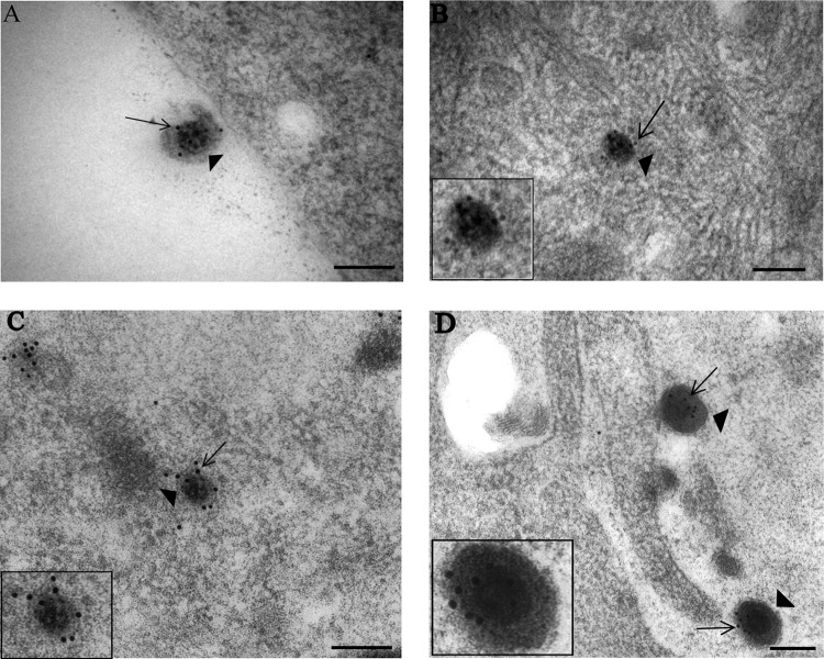
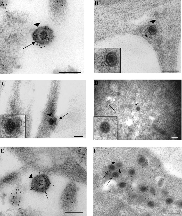
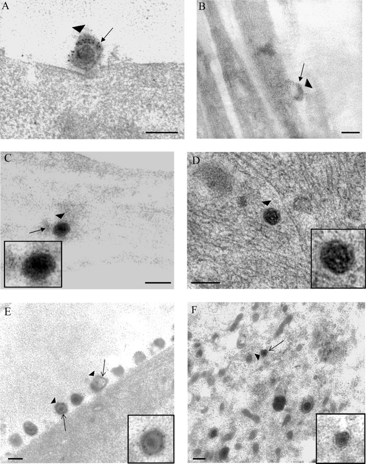
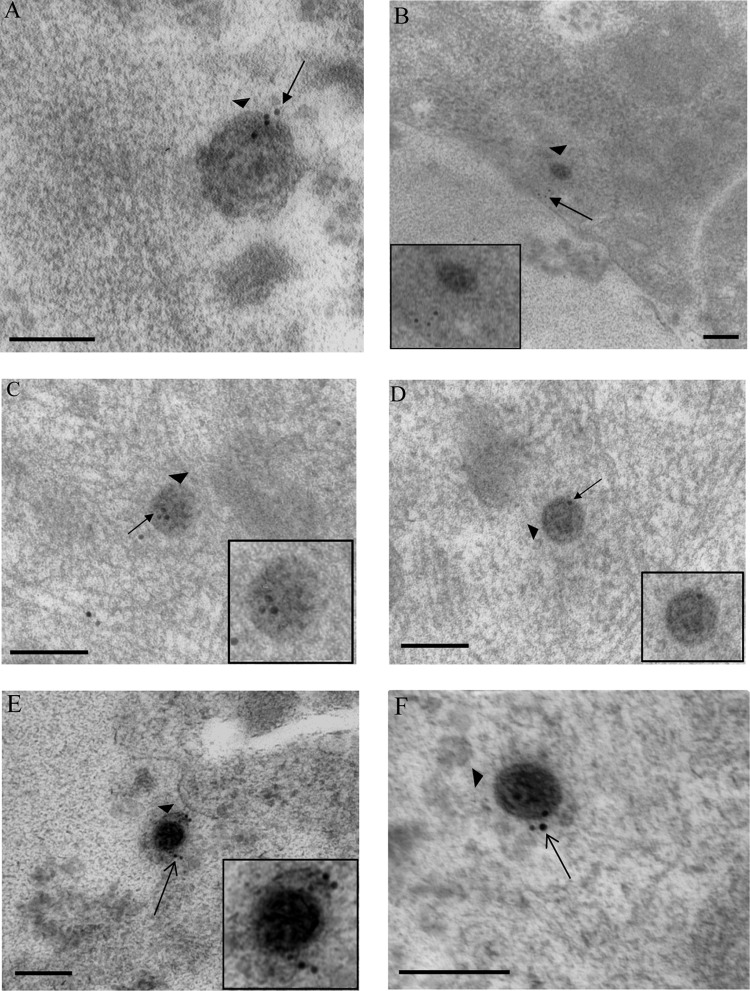
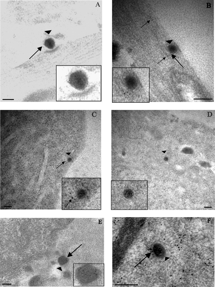
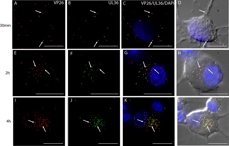
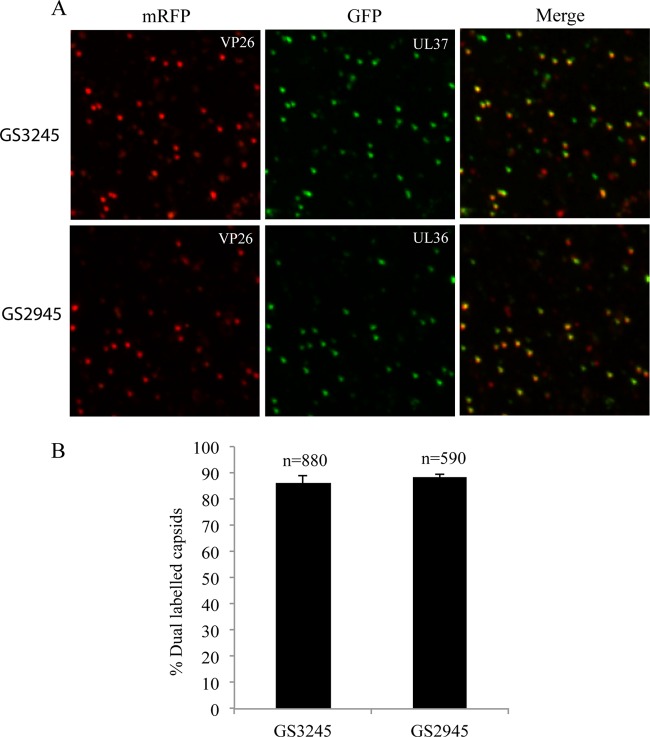
Herpes simplex virus 1 (HSV-1) enters neurons primarily by fusion of the viral envelope with the host cell plasma membrane, leading to the release of the capsid into the cytosol. The capsid travels via microtubule-mediated retrograde transport to the nuclear membrane, where the viral DNA is released for replication in the nucleus. In the present study, the composition and kinetics of incoming HSV-1 capsids during entry and retrograde transport in axons of human fetal and dissociated rat dorsal root ganglia (DRG) neurons were examined by wide-field deconvolution microscopy and transmission immunoelectron microscopy (TIEM). We show that HSV-1 tegument proteins, including VP16, VP22, most pUL37, and some pUL36, dissociated from the incoming virions. The inner tegument proteins, including pUL36 and some pUL37, remained associated with the capsid during virus entry and transit to the nucleus in the neuronal cell body. By TIEM, a progressive loss of tegument proteins, including VP16, VP22, most pUL37, and some pUL36, was observed, with most of the tegument dissociating at the plasma membrane of the axons and the neuronal cell body. Further dissociation occurred within the axons and the cytosol as the capsids moved to the nucleus, resulting in the release of free tegument proteins, especially VP16, VP22, pUL37, and some pUL36, into the cytosol. This study elucidates ultrastructurally the composition of HSV-1 capsids that encounter the microtubules in the core of human axons and the complement of free tegument proteins released into the cytosol during virus entry.


Figures

Fig 1
Fig 1
Immunogold labeling for capsid VP5 of HSV-1 particles in vUL37-GFP-infected human axons and rat DRG neurons. Coverslips with DRG cultures were processed with Lowicryl HM20, and immunogold labeling for VP5 of ultrathin sections was performed as described in Materials and Methods. (A) Extracellular virion (arrowhead) with label for VP5 (arrow) and lying close to a human DRG axon at 30 mpi. (B) Unenveloped capsid (arrowhead) within the cytosol of the cell body of a rat DRG neuron, labeled for VP5 (arrow), at 4 hpi. (C) Unenveloped capsid (arrowhead) within a neuronal cell body of a rat DRG neuron, labeled for VP5 (arrow), at 24 hpi. (D) Extracellular virions (arrowheads), labeled for VP5 (arrows) and lying close to a human axon at 24 hpi. (Insets) Enlargements of capsids in each panel. NM, nuclear membrane; PM, plasma membrane. Bars, 200 nm.
Fig 2
Fig 2
Immunogold labeling for tegument protein VP16 in human axons and rat neurons infected with vUL37-GFP. (A) Extracellular virion (arrowhead) lying close to a human axon and carrying label for VP16 (arrow) at 30 mpi. (B and C) Unenveloped capsids (arrowheads) within human axons at 30 mpi. Label for VP16 (arrows) is present off the capsids and in panel C is on the plasma membrane. (D) Unenveloped capsid with no label for VP16, present in the cytosol of cell body of a rat DRG neuron at 2 hpi. Diffuse free label for VP16 is present in the cytosol (arrow). (E) Extracellular virion (arrowhead), labeled for VP16 (arrow), in close proximity to the plasma membrane of a DRG neuron at 24 hpi. (F) Unenveloped cytoplasmic capsids (arrowheads) inside the cytosol of the cell body of a rat DRG neuron at 24 hpi, carrying label for VP16 (arrows). (Insets) Enlargements of capsids in each panel. Gold particles were 5 nm (B and C) or 10 nm (other panels). Bars, 200 nm.
Fig 3
Fig 3
Immunogold labeling for tegument protein VP22 in human axons and rat neurons infected with vUL37-GFP. Extracellular virions (arrowheads) bound to the plasma membrane of a rat DRG neuron (A) and human DRG axons (B) at 30 mpi. Label for VP22 is present on the virions (arrows). (C) Unenveloped capsid (arrowhead) within a human DRG axon at 30 mpi. Label for VP22 is off the capsid. (Inset) Enlargement of the capsid. (D) Unenveloped capsid (arrowhead) in the cytosol of cell body of a rat DRG neuron at 4 hpi with no label for VP22. (Inset) Enlargement of the capsid. (E) Extracellular virions (arrowheads) lining the cell surface at 24 hpi and labeled for VP22 (arrows). (Inset) Enlargement of a virion. (F) Unenveloped capsid (arrowhead) in the cytosol of the cell body of a rat DRG neuron carrying label for VP22 (arrow) at 24 hpi. (Inset) Enlargement of a capsid. Gold particles were 5 nm (C) or 10 nm (other panels). Bars, 200 nm.
Fig 4
Fig 4
Immunogold labeling for tegument pUL36 in human axons and rat DRG neurons infected with vUL37-GFP. (A) Extracellular virion (arrowhead), labeled for pUL36 (arrow), bound to the plasma membrane of the cell body of rat DRG neuron at 30 mpi. (B) Unenveloped capsid (arrowhead) in the cytosol of the cell body of a rat DRG neuron at 30 mpi. Label for pUL36 (arrow) is off the viral capsid. (C and D) Unenveloped capsids (arrowheads) in the cytosol of cell body of rat DRG neurons, carrying label for pUL36 (arrows) at 2 hpi and 4 hpi, respectively. (E and F) Extracellular virus (arrowhead) (E) and unenveloped capsid (arrowhead) (F) in the cytosol of cell body of a rat DRG neuron, carrying label for pUL36 (arrows), at 24 hpi. (Insets) Enlargements of viral particles in each panel. Bars, 200 nm.
Fig 5
Fig 5
Immunogold labeling for tegument protein pUL37 in human axons and rat DRG neurons infected with vUL37-GFP. (A) Extracellular virion (arrowhead), labeled for pUL37 (arrow), bound to the plasma membrane of a human DRG axon at 30 mpi. (B) Unenveloped capsid (arrowhead), labeled for pUL37 (long arrow), adjacent to the inner aspect of the plasma membrane of a human DRG axon at 30 mpi. Label for pUL37 is both on (long arrow) and off (short arrows) the capsid. (C) Unenveloped capsid (arrowhead) in the cytosol of cell body of a rat DRG neuron at 2 hpi, lying close to the plasma membrane and with label (arrow) off the capsid. (D) Unenveloped capsid (arrowhead) present deep in the cytosol of the cell body of a rat DRG neuron at 4 hpi and carrying no label for pUL37. (E and F) Extracellular virion (E) and cytoplasmic unenveloped capsid (F) (arrowheads) labeled for pUL37 (arrows) at 24 hpi. (Insets) Enlargements of virions in each panel. Bars, 200 nm.
Fig 6
Fig 6
Visualization of mRFP1 capsids and pUL36-GFP in rat DRG neurons infected with HSV F-GS2945 from 30 mpi to 4 hpi by wide-field deconvolution microscopy. The images are three-dimensional (3D) reconstructed z series, and areas of colocalization are in yellow. Fluorescent puncta representing mRFP1 capsids (red) (A) and pUL36-GFP (green) (B) were detected at the cell periphery and along axons at 30 mpi (arrows). Most of the mRFP1 capsids colocalized with pUL36-GFP at the cell periphery and along axons at this time (C and D, arrows). By 2 hpi, fluorescent puncta for mRFP1 capsids (E) and for pUL36-GFP (F) were detected in the cytosol of the cell body and at the nuclear rim (arrows). The majority of mRFP1 capsids in the cytosol (78.9%; n = 71) and at the nuclear rim (71.2%; n = 59) colocalized with pUL36-GFP (G and H; arrows). Similarly, at 4 hpi, the majority of the fluorescent puncta for mRFP1 capsids (I) present in the cytosol (71.3%; n = 237) and at the nuclear rim (70.3%; n = 202) colocalized with fluorescent puncta (J) for pUL36-GFP (I to L; arrows). Bars, 10 μm.
Fig 7
Fig 7
Visualization of mRFP1 capsids and GFP-pUL37 in rat DRG neurons infected with HSV F-GS3245 from 30 mpi to 4 hpi by wide-field deconvolution microscopy. These images are 3D reconstructed z series, and areas of colocalization appear in yellow. Fluorescent puncta representing mRFP1 capsids (red) (A) and GFP-pUL37 (green) (B) were detected at the cell periphery and along axons at 30 mpi (arrows). Most of the mRFP1 capsids colocalized with GFP-pUL37 at the cell periphery and along axons at this time (C and D; arrows). By 2 hpi, fluorescent puncta for mRFP1 capsids (E and I) and for GFP-pUL37 (F and J) were detected in the cytosol of the cell body and at the nuclear rim (arrows). About half of the mRFP1 capsids in the cytosol (55.2%; n = 67) and at the nuclear rim (51.3%; n = 76) colocalized with GFP-pUL37 (G, H, K, and L; arrows). By 4 hpi, less than half of fluorescent puncta for mRFP1 capsids (M) present in the cytosol (38.3%; n = 162) and at the nuclear rim (37.2%; n = 180) colocalized with fluorescent puncta (N) for GFP-pUL37 (O and P) (arrows). Bars, 10 μm.
Fig 8
Fig 8
Characterization of F-GS2945 and F-GS3245 extracellular virus particles. (A) Cell supernatants containing extracellular virus particles were spun down on Cell Tak-coated glass coverslips at 1,200 × g for 30 min at 4°C followed by fixation in 3% formaldehyde and imaged by deconvolution microscopy. F-GS3245 expresses mRFP1-VP26 and GFP-pUL37, while F-GS2945 expresses mRFP1-VP26 and pUL36-GFP. “Merge” panels show mRFP fluorescence superimposed on GFP fluorescence. (B) Percentage of mRFP capsids emitting GFP fluorescence from supernatants of F-GS3245 and F-GS2945 shown in panel A. Results are averages from five separate counts. Error bars represent standard errors of means. n, total number of capsids.

References

    1. Akhtar J, Shukla D. 2009. Viral entry mechanisms: cellular and viral mediators of herpes simplex virus entry. FEBS J. 276:7228–7236 - PMC - PubMed
    1. Amici C, et al. 2006. Herpes simplex virus disrupts NF-κB regulation by blocking its recruitment on the IκBα promoter and directing the factor on viral genes. J. Biol. Chem. 281:7110–7117 - PubMed
    1. Antinone SE, Smith GA. 2010. Retrograde axon transport of herpes simplex virus and pseudorabies virus: a live-cell comparative analysis. J. Virol. 84:1504–1512 - PMC - PubMed
    1. Antinone SE, Zaichick SV, Smith GA. 2010. Resolving the assembly state of herpes simplex virus during axon transport by live-cell imaging. J. Virol. 84:13019–13030 - PMC - PubMed
    1. Batterson W, Furlong D, Roizman B. 1983. Molecular genetics of herpes simplex virus. VIII. further characterization of a temperature-sensitive mutant defective in release of viral DNA and in other stages of the viral reproductive cycle. J. Virol. 45:397–407 - PMC - PubMed

Publication types

Substances

LinkOut - more resources