. 2012;8(3):e1002584.
doi: 10.1371/journal.ppat.1002584. Epub 2012 Mar 22.

Dengue virus infection perturbs lipid homeostasis in infected mosquito cells

Affiliations

Dengue virus infection perturbs lipid homeostasis in infected mosquito cells

Rushika Perera et al. PLoS Pathog. 2012.

Abstract

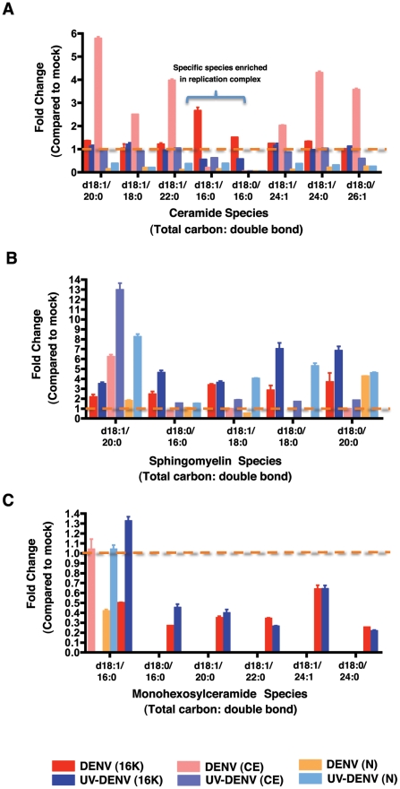
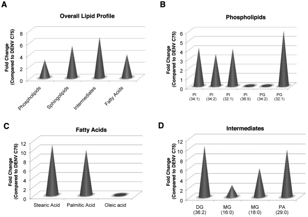
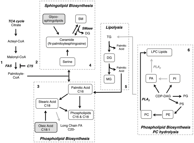
Dengue virus causes ∼50-100 million infections per year and thus is considered one of the most aggressive arthropod-borne human pathogen worldwide. During its replication, dengue virus induces dramatic alterations in the intracellular membranes of infected cells. This phenomenon is observed both in human and vector-derived cells. Using high-resolution mass spectrometry of mosquito cells, we show that this membrane remodeling is directly linked to a unique lipid repertoire induced by dengue virus infection. Specifically, 15% of the metabolites detected were significantly different between DENV infected and uninfected cells while 85% of the metabolites detected were significantly different in isolated replication complex membranes. Furthermore, we demonstrate that intracellular lipid redistribution induced by the inhibition of fatty acid synthase, the rate-limiting enzyme in lipid biosynthesis, is sufficient for cell survival but is inhibitory to dengue virus replication. Lipids that have the capacity to destabilize and change the curvature of membranes as well as lipids that change the permeability of membranes are enriched in dengue virus infected cells. Several sphingolipids and other bioactive signaling molecules that are involved in controlling membrane fusion, fission, and trafficking as well as molecules that influence cytoskeletal reorganization are also up regulated during dengue infection. These observations shed light on the emerging role of lipids in shaping the membrane and protein environments during viral infections and suggest membrane-organizing principles that may influence virus-induced intracellular membrane architecture.


Conflict of interest statement

The authors have declared that no competing interests exist.

Figures

Figure 1
Figure 1. C75, a fatty acid synthase inhibitor disrupts DENV replication in mosquito cells.
A. C6/36 mosquito cells were infected with DENV at an MOI of 3. Following adsorption of the virus, the indicated concentrations of C75 were added to the cells in the overlay media. A vehicle control of ethanol only was also included. Virus release was assayed by plaque assay 24 hr post-infection. Cell viability was assayed using the Quick Cell Proliferation Kit (Promega) at 24 hr post-infection. B. A time course of addition of C75 in C6/36 cells infected with DENV at an MOI of 3. Cells were either pre-treated (2 hr), or treated with 6.3 uM C75 during adsorption, post-adsorption (0 hr) or at 4, 8 and 12 hr post-infection. The results represent three independent experiments. The error bars represent standard deviation of the mean.
Figure 2
Figure 2. A plot of the principle components analysis scores shows segregation of the global lipid profile between uninfected and DENV-infected mosquito cells.
The abundance of lipids in C6/36 cells infected with either DENV (MOI 20) or UV-DENV was measured at 36 and 60 hr post-infection and compared to uninfected controls (mock). Each experiment included 4 independent replicates. A total of 7217 features were compared by principle component analysis (PCA). The plot shows differences that are specific to infectious virus (DENV), a non-replicating virus (DENV-UV) and the mock control.
Figure 3
Figure 3. Whole cell lipidomics reveal an altered lipid composition in DENV infected cells.
Panel A and B represent an average expression (fold change) of the total number of individual lipids significantly expressed (p<0.05) per lipid class at 36 and 60 hr post-infection, respectively. The fold changes represent DENV-infected cells or UV-DENV exposed cells compared to the mock control. A lack of cones indicates that the expression level of those specific lipids were not significant (p<0.05). Panels C–H are representative lipid molecular species from specific lipid classes significantly regulated at the two different time points. The data are plotted as the integrated LC-MS peak abundance, in log 2 scale with standard deviation. PC, phosphatidylcholine; PE, phosphatidylethanolamine; SM, sphingomyelin; CER, ceramide; CER-PE, ceramide phosphoethanolamine; Lyso, lysophospholipids. See supplementary table S1 for a complete list of lipid features detected in this study. Four replicates were included in the lipidomic analyses. The error bars represent standard deviation of the mean. The blue dashed line separates species that remain elevated at both time points (36 and 60 hr) from species that are only elevated at the 36 hr time point. Infections were carried out using an MOI of 20 in C6/36 cells. Significantly expressed lipids species are shown denoted with an asterisk (*).
Figure 4
Figure 4. Newly synthesized lipids and viral RNA in subcellular fractions show a dynamic distribution with time of infection.
A. A pulse-chase analysis of 14C-acetate incorporation into newly synthesized lipids. The results show total labeled lipid in post-nuclear supernatants of C6/36 cells infected with DENV for 36 and 60 hr at an MOI of 20. B. The same pulse-chase analysis showing 14C-acetate incorporation into newly synthesized lipids in subcellular fractions (16K and CE) at 36 and 60 hr post-infection. C. The ratio of viral RNA genome copies per labeled lipid in subcellular fractions (described in B) at 36 and 60 hr post-infection. 16K, membrane fraction (pellet) following centrifugation of post-nuclear supernatants at 16, 000× g. CE, cytoplasmic extract following centrifugation of post-nuclear supernatants at 16,000× g. cpm, counts per minute.
Figure 5
Figure 5. Several lipid classes are differentially regulated in replication complex membranes isolated from DENV infected cells.
Panel A shows fold changes for each lipid class significantly (p<0.05) regulated in membrane fractions isolated from DENV infected (red) or UV-DENV exposed (blue) cells compared to the mock control. Infection was carried out using an MOI of 20. Panels B–D show fold changes for individual molecular species within each lipid class that are regulated. PC, phosphatidylcholine; PE, phosphatidylethanolamine; PG, phosphatidylglycerol; MG, monoacylglycerol; DG, diacylglycerol; PA, phosphatidic acid. See supplementary table S2 for a complete list of lipid features detected in this analysis and supplementary figure 1 for a heat map representation of the data. Three replicates were included in the lipidomic analyses.
Figure 6
Figure 6. Bioactive sphingolipid species are differentially regulated in replication complex membranes isolated from DENV-infected mosquito cells.
Multiple Reaction Monitoring (MRM) analysis of sphingolipids species differentially regulated in DENV-infected cells (MOI 20) or UV-DENV exposed cells compared to the mock control (see also supplementary table S3). The data represent fold changes observed in three subcellular fractions that were analyzed in this study; 16K, replication complex membranes; CE, cytoplasmic extracts following removal of replication complex membranes and nuclei; N, nuclear fraction. Panels A–C represent ceramide, sphingomyelin and monohexosylceramide species respectively. The dashed line highlights values equal to the mock. The data represent three independent experiments. The error bars represent standard deviation of the mean.
Figure 7
Figure 7. The Lipid repertoire of DENV infected mosquito cells is unfavorable for replication in the presence of C75.
The specific lipid classes differentially regulated by C75 treatment of cells are shown. Panel A represents the fold changes of lipid classes expressed in DENV infected cells (MOI 3) compared to C75 treated DENV infected cells. Panels B–D show fold changes for individual molecular species within each lipid class that are regulated. PI, phosphatidylinositol; PG, phosphatidylglycerol; MG, monoacylglycerol; DG, diacylglycerol; PA, phosphatidic acid. See supplementary table S3 for a complete list of lipid features detected in this analysis and supplementary figure 2 for a heat map representation of the data. Three replicates were included in the lipidomic analyses.
Figure 8
Figure 8. Dengue virus infection perturbs lipid homestasis in infected mosquito cells.
The lipidomic analyses of dengue virus infected C6/36 mosquito cells suggest several metabolic pathways that may be significantly up regulated during infection. The grey dashed line highlights specific pathways of interest. Black arrows highlight reactions suggested by the lipidomic data and grey arrows represent reactions not observed in the data. Metabolites highlighted in boxes (solid line) are up regulated (white) or down regulated (grey) in DENV infected mosquito cells. 1. Through the recruitment and activation of FAS, DENV stimulates de novo phospholipid biosynthesis in the replication complex . 2. Inhibition of this process with C75 disrupts the cellular lipid repertoire in mosquito cells to be unfavorable for virus replication. 3. The lipidomic analyses reveal an up regulation of fatty acids such as palmitic (C16) and stearic (C18) acid. These fatty acids are intermediates in the biosynthesis of phospholipids, which is up regulated during DENV infection. Interestingly, in DENV infected cells the prevalent phospholipids primarily consist of C16 and C18 unsaturated acyl chains. Very long chain fatty acids are not significantly up regulated during infection. 4. FAS activity also stimulates de novo sphingolipid biosynthesis. In the lipidomic analyses, the up regulation of intermediates such as N-palmitoylesphingosine suggests sphingolipid biosynthesis is activated during DENV infection. Specifically, SM and CER are enriched in DENV infected cells. Alternately, the up regulation in CER (and DG) during infection could result from the degradation of SM through the activity of sphingomyelinases (Smase). The resulting CER and DG could be redirected into several signaling pathways or be utilized for de novo phospholipid biosynthesis. The glycopshingolipids, GlcCER and GalCER are down regulated during DENV infection, which suggest that they are catabolized to produce CER. 5. Lipidomic analyses also suggest the up regulation of triacylglycerol catabolism (Lipolysis) in DENV infected cells. This pathway results in the generation of MG, DG and palmitic acid. These intermediates are all up regulated in DENV infected cells and could be utilized for downstream signaling or de novo phospholipid biosynthesis. It has also been shown that TG catabolism is necessary for mitochondrial β-oxidation during DENV infection . 6. Elevated levels of LPC in DENV infected cells also suggest activation of PC hydrolysis by PLA2. This enzyme is activated during DENV infection. The elevated levels of other phospholipids such as PA, PI, PE, PG as well as PC suggest that the CDG-DG pathway for phospholipid biosynthesis could also be activated. FAS, fatty acid synthase; DENV, dengue virus; C75, inhibitor of FAS; SM, sphingomyelin; CER, ceramide; MG, monoacylglycerol; DG, diacylglycerol; TG, triacylglycerol; LPC, lysophosphatidylcholine; PLA2, phospholipase A2; PA, phosphatidic acid; PI, phosphatidylinositol; PE, phosphatidylethanolamine; PG, phosphatidylglycerol; PC, phosphatidylcholine.

References

    1. Visser B, Le Lann C, den Blanken FJ, Harvey JA, van Alphen JJ, et al. Loss of lipid synthesis as an evolutionary consequence of a parasitic lifestyle. Proc Natl Acad Sci U S A. 2010;107:8677–82. - PMC - PubMed
    1. Diaz A, Wang X, Ahlquist P. Membrane-shaping host reticulon proteins play crucial roles in viral RNA replication compartment formation and function. Proc Natl Acad Sci U S A. 2010;107:16291–6. - PMC - PubMed
    1. Gillespie LK, Hoenen A, Morgan G, Mackenzie JM. The endoplasmic reticulum provides the membrane platform for biogenesis of the flavivirus replication complex. J Virol. 2010;84:10438–47. - PMC - PubMed
    1. Welsch S, Miller S, Romero-Brey I, Merz A, Bleck CK, et al. Composition and three-dimensional architecture of the dengue virus replication and assembly sites. Cell Host Microbe. 2009;5:365–75. - PMC - PubMed
    1. Jackson WT, Giddings TH, Jr, Taylor MP, Mulinyawe S, Rabinovitch M, et al. Subversion of cellular autophagosomal machinery by RNA viruses. PLoS Biol. 2005;3:156. - PMC - PubMed

Publication types

MeSH terms

Substances