. 2012;8(3):e1002601.
doi: 10.1371/journal.ppat.1002601. Epub 2012 Mar 22.

Direct recognition of Fusobacterium nucleatum by the NK cell natural cytotoxicity receptor NKp46 aggravates periodontal disease

Affiliations

Direct recognition of Fusobacterium nucleatum by the NK cell natural cytotoxicity receptor NKp46 aggravates periodontal disease

Stella Chaushu et al. PLoS Pathog. 2012.

Abstract

Periodontitis is a common human chronic inflammatory disease that results in the destruction of the tooth attachment apparatus and tooth loss. Although infections with periopathogenic bacteria such as Porphyromonas gingivalis (P. gingivalis) and Fusobacterium nucleatum (F. nucleatum) are essential for inducing periodontitis, the nature and magnitude of the disease is determined by the host's immune response. Here, we investigate the role played by the NK killer receptor NKp46 (NCR1 in mice), in the pathogenesis of periodontitis. Using an oral infection periodontitis model we demonstrate that following F. nucleatum infection no alveolar bone loss is observed in mice deficient for NCR1 expression, whereas around 20% bone loss is observed in wild type mice and in mice infected with P. gingivalis. By using subcutaneous chambers inoculated with F. nucleatum we demonstrate that immune cells, including NK cells, rapidly accumulate in the chambers and that this leads to a fast and transient, NCR1-dependant TNF-α secretion. We further show that both the mouse NCR1 and the human NKp46 bind directly to F. nucleatum and we demonstrate that this binding is sensitive to heat, to proteinase K and to pronase treatments. Finally, we show in vitro that the interaction of NK cells with F. nucleatum leads to an NCR1-dependent secretion of TNF-α. Thus, the present study provides the first evidence that NCR1 and NKp46 directly recognize a periodontal pathogen and that this interaction influences the outcome of F. nucleatum-mediated periodontitis.


Conflict of interest statement

The authors have declared that no competing interests exist.

Figures

Figure 1
Figure 1. NCR1-dependent bone loss.
(A) Mesio-distal view on a random section. Red, yellow, gray and green represent the enamel, dentin, bone and the residual bone area above the reference line, respectively. (B) Ncr1+/+ (WT) and Ncr1gfp/gfp (KO) mice (n = 8 in each group) were challenged orally three times at 2-day intervals with inoculums of F. nucleatum and of P. gingivalis (2×10⁸ CFU in 0.2 ml of PBS and 2% carboxymethylcellulose). Six weeks later, the jaws were harvested and the residual alveolar bone volume (mm3×10−3) was measured. Error bars represent SD. Data represent percentage of bone loss ± SD and are average of two independent experiments. ** p<0.01. The difference in bone loss between the KO and the WT mice following P. gingivalis inoculation was not statistically significant, p<0.09.
Figure 2
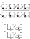
Figure 2. Infiltration of cells into challenged chambers.
Immune cells in the chambers were analyzed by FACS 2 hours (A) and 24 hours (B) following bacteria inoculation. The intrinsic GFP labeling marks mainly NK cells. Staining was performed with Phycoerythrin conjugated anti-DX5 and anti-NKG2D mAbs. Staining with NK1.1 was visualized by using Cy-5 conjugated strepavidin. The cell percentage and cell numbers is indicated in each quadrant. One representative experiment out of four is shown.
Figure 3
Figure 3. Survival of bacteria in chamber exudates.
Viable CFU from chamber exudates were determined, 2 h and 24 h following inoculation of P. gingivalis, F. nucleatum or E. coli. Each symbol represents an individual mouse. One representative experiment out of four is shown. The y axis represents number of colonies (logarithmic scale).
Figure 4
Figure 4. Cytokine profiles in challenged chambers.
TNF-α levels were determined by ELISA in chamber exudates of Ncr1+/+ (WT) and Ncr1gfp/gfp (KO) mice at baseline (0), 2 h and 24 h following bacterial challenge. Data represent absorbance at 650 nm ± SD and are average of three different experiments. ** p<0.01.
Figure 5
Figure 5. The infiltration of immune cells into the F. nucleatum challenged chambers is NCR1-independent.
The presence of immune cells; (A) lymphocytes and (B) DC (identified by CD11c) and macrophages (identified by F4/80) in the chambers were analyzed by FACS, 2 hours following the injection of F. nucleatum. The mAb used for staining is indicated in the Y axis. IC is isotype control. The percentage of the various cells and the cell numbers are indicated in each quadrant. One representative experiment out of four is shown.
Figure 6
Figure 6. In vivo response to heat-treated F. nucleatum bacteria.
(A) FACS analysis of chamber exudates, 2 hours and 24 hours following challenge with heat-killed F. nucleatum. Staining was performed with Phycoerythrin conjugated anti-DX5 mAb. The percentages of GFP+ NK cells are indicated. One representative experiment out of three is shown. (B) TNF-α level in chamber exudates of Ncr1+/+ (WT) and Ncr1gfp/gfp (KO) was determined by ELISA, 2 and 24 hours following challenge with viable and heat-killed F. nucleatum. Data represent absorbance at 650 nm ± SD and are average of two different experiments. * p<0.05, ** p<0.01.
Figure 7
Figure 7. Direct binding of NCR1 and NKp46 to F. nucleatum.
F. nucleatum (A), P. gingivalis (B), uninfected 721.221 cells (C), and 721.221 cells infected with influenza PR8 virus (D), were stained with NCR1-Ig (left) and with NKp46-Ig (right) at two different dosages; 5 µg, blue line and 1 µg, red line. Staining was visualized using a Phycoerythrin conjugated anti-human Ig antibody. The filled grey histograms represent control staining with the NKp46D1-Ig fusion protein. The Median Fluorescence Intensity (MFI) values of the D1-Ig, NCR1-Ig and NKp46 staining are indicated in each of the histograms. One representative experiment out of five is shown. (E) F. nucleatum (upper) and P. gingivalis (lower) bacteria were stained with various activating NK cell receptors fused to Ig (indicated above the quadrants). Figure shows one representative experiment out of three performed.
Figure 8
Figure 8. The F. nucleatum ligand is sensitive to heat, proteinase K and pronase treatment.
Viable untreated, or treated (treatments are indicated above the histograms) F. nucleatum bacteria were stained with 5 µg of NCR1-Ig (upper) and of NKp46-Ig (lower). Filled grey histograms represent the staining with the NKp46D1-Ig. The blue histograms represent the staining of the untreated F. nucleatum and the red histograms represent the staining of the treated F. nucleatum. One representative experiment out of three is shown.
Figure 9
Figure 9. Functional reporter assays.
(A) Basal IL-2 secretion of BW-NCR1, BW-NKp46 and parental BW cells. (B) BW-NKp46 transfected cells were cultured for 48 h in presence of plate bound (dosages indicated in the X axis) anti NKp46 mAb and control mAb. (C) 5×10⁴ BW-NKp46 transfected cells were cultured for 48 hours, with treated or untreated F. nucleatum and with 721.221 cells infected with PR8 influenza virus in the presence and in the absence of anti-NKp46 mAb. IL-2 secretion was determined by ELISA. Data represent absorbance at 650 nm ± SD and are average of two different experiments * p<0.05, ** p<0.01.
Figure 10
Figure 10. NCR1-dependent secretion of TNF-α following interaction with F. nucleatum.
NK cells isolated from Ncr1+/gfp (Het) and from Ncr1gfp/gfp (KO) mice were cultured with F. nucleatum and with P. gingivalis bacteria for 48 hours. TNF-α secretion was determined by ELISA. Data represent absorbance at 650 nm ± SD and are average of two different experiments. *** p<0.001.

References

    1. Albandar JM, Brunelle JA, Kingman A. Destructive periodontal disease in adults 30 years of age and older in the United States, 1988–1994. J Periodontol. 1999;70:13–29. - PubMed
    1. Wu T, Trevisan M, Genco RJ, Dorn JP, Falkner KL, et al. Periodontal disease and risk of cerebrovascular disease: the first national health and nutrition examination survey and its follow-up study. Arch Intern Med. 2000;160:2749–2755. - PubMed
    1. Scannapieco FA. Role of oral bacteria in respiratory infection. J Periodontol. 1999;70:793–802. - PubMed
    1. Offenbacher S, Jared HL, O'Reilly PG, Wells SR, Salvi GE, et al. Potential pathogenic mechanisms of periodontitis associated pregnancy complications. Ann Periodontol. 1998;3:233–250. - PubMed
    1. Dodman T, Robson J, Pincus D. Kingella kingae infections in children. J Paediatr Child Health. 2000;36:87–90. - PubMed

Publication types

MeSH terms

Substances

LinkOut - more resources