. 2012;7(3):e33241.
doi: 10.1371/journal.pone.0033241. Epub 2012 Mar 22.

Towards endometriosis diagnosis by gadofosveset-trisodium enhanced magnetic resonance imaging

Affiliations

Towards endometriosis diagnosis by gadofosveset-trisodium enhanced magnetic resonance imaging

Marc H Schreinemacher et al. PLoS One. 2012.

Abstract

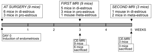
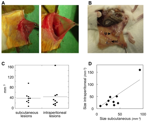
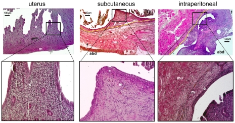
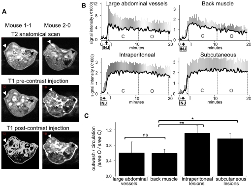
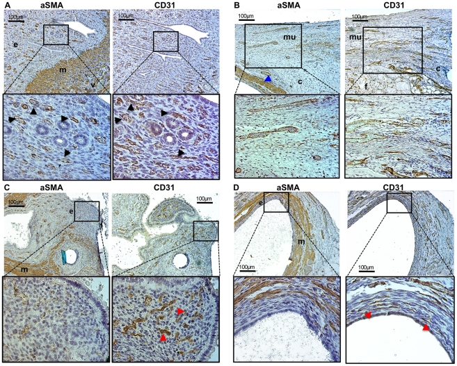
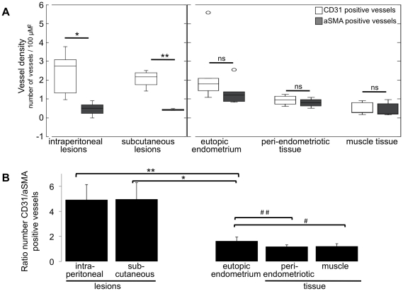
Endometriosis is defined as the presence of endometrial tissue outside the uterus. It affects 10-15% of women during reproductive age and has a big personal and social impact due to chronic pelvic pain, subfertility, loss of work-hours and medical costs. Such conditions are exacerbated by the fact that the correct diagnosis is made as late as 8-11 years after symptom presentation. This is due to the lack of a reliable non-invasive diagnostic test and the fact that the reference diagnostic standard is laparoscopy (invasive, expensive and not without risks). High-molecular weight gadofosveset-trisodium is used as contrast agent in Magnetic Resonance Imaging (MRI). Since it extravasates from hyperpermeable vessels more easily than from mature blood vessels, this contrast agent detects angiogenesis efficiently. Endometriosis has high angiogenic activity. Therefore, we have tested the possibility to detect endometriosis non-invasively using Dynamic Contrast-Enhanced MRI (DCE-MRI) and gadofosveset-trisodium as a contrast agent in a mouse model. Endometriotic lesions were surgically induced in nine mice by autologous transplantation. Three weeks after lesion induction, mice were scanned by DCE-MRI. Dynamic image analysis showed that the rates of uptake (inwash), persistence and outwash of the contrast agent were different between endometriosis and control tissues (large blood vessels and back muscle). Due to the extensive angiogenesis in induced lesions, the contrast agent persisted longer in endometriotic than control tissues, thus enhancing the MRI signal intensity. DCE-MRI was repeated five weeks after lesion induction, and contrast enhancement was similar to that observed three weeks after endometriosis induction. The endothelial-cell marker CD31 and the pericyte marker α-smooth-muscle-actin (mature vessels) were detected with immunohistochemistry and confirmed that endometriotic lesions had significantly higher prevalence of new vessels (CD31 only positive) than the uterus and control tissues. The diagnostic value of gadofosveset-trisodium to detect endometriosis should be tested in human settings.


Conflict of interest statement

Competing Interests: The authors have declared that no competing interests exist.

Figures

Figure 1
Figure 1. Scheme of the animal experiment.
Endometriosis was surgically induced by autologous transplantation in nine mice at DAY 0. Three weeks (all nine mice) and five weeks later (three mice), animals were subjected to DCE-MRI.
Figure 2
Figure 2. Endometriosis was induced in mice.
A. Autologous transplantation : one uterine horn from one mouse is removed, opened longitudinally, and two 2–3 mm2 uterine fragments are sutured to the abdominal wall of the same mouse either subcutaneously (left) or intraperitoneally (right). B. At sacrifice, macroscopic inspection revealed the presence of both subcutaneous (sc) and intraperitoneal (ip) lesions. C. No significant size differences were seen between subcutaneous and intraperitoneal lesions. D. The size of the subcutaneous and intraperitoneal lesions in each single mouse are plotted on the X and Y-axes, respectively, and showed a significant correlation (R2 = 0.7930).
Figure 3
Figure 3. Endometriosis was confirmed with histology.
Haematoxylin and eosin staining was used to characterise histologically the lesions. ‘e’ indicates endometrial tissue; ‘m’ = myometrium. Muscle fibres and fat tissue of the abdominal wall are indicated by: ‘mu’ = muscle; ‘f’ = fat; ‘abd’ = abdominal cavity. The border between endometriosis and the abdominal wall is indicated by the yellow-dashed line.
Figure 4
Figure 4. Endometriosis visualised by gadofosveset-trisodium enhanced MRI.
Three weeks after lesion induction, mice were subjected to gadofosveset-trisodium enhanced MRI. A. Examples of T2 weighted anatomical images and T1 weighted prior and post contrast injection in two mice. Lesions are indicated by black arrow-heads (subcutaneous) and white arrow-heads (intraperitoneal). B. DCE-MRI derived intensity time-curves in the large abdominal blood vessels, back muscle, intraperitoneal and subcutaneous lesions. Injection of gadofosveset-trisodium is indicated (arrow-INJ). The inwash (I), circulation (C) and the outwash phases (O) of the contrast agent are indicated. Charts represent the average signal intensity of nine mice plus SD. C. The ratio O/C was assessed in the large abdominal vessels, back muscle and endometriosis. Statistic based on unpaired Wilcoxon test: * = 0.001; **<0.001; ns = non-significant.
Figure 5
Figure 5. Endometriosis is characterised by angiogenesis.
Different tissues were stained by immunohistochemistry for CD31 (endothelial cell marker) and the pericyte marker aSMA (present in mature vessels only). Representative images are shown. A. In the uterus (eutopic endometrium), most CD31 positive blood vessels are also aSMA positive, indicated by the black arrow-heads in the enlargements. ‘e’ = endometrium; ‘m’ = aSMA positive myometrial cells; ‘v’ = large-mature blood vessels present in the myometrial layer. B. Abdominal wall consisting in large part of striated muscular tissue (mu), with some connective (c) and fat (f) tissues. A surgically induced endometriotic lesion is present on the left image, and visible as aSMA positive myometrial tissue (at the bottom-left of the low-magnification image; blue arrow-head). C & D. Intraperitoneal (C) and subcutaneous (D) lesions. In the endometrial layer of the lesions (e), most blood vessels are DC31 only positive (red arrow-heads in the enlargements); ‘m’ = myometrium (aSMA positive cells).
Figure 6
Figure 6. Quantification of new and mature blood vessels.
A. The number of CD31 and aSMA positive blood vessels, or vessel density (number vessels per 100 µm2) was quantified in: intraperitoneally and subcutaneously induced endometriosis; eutopic endometrium (inside the uterus); muscular tissue of the abdominal wall either adjacent (peri-endometriotic) or distant to the induced lesions (muscle). The average number of vessels in each tissue for six mice is shown; statistic is based on unpaired Wilcoxon test: * = 0.002; ** = 0.029; ns = non-significantly different. B. The ratio between CD31 and aSMA positive blood vessels was calculated in corresponding locations (endometriosis, uterus, peri-endometriosis and muscle) in each mouse. Afterwards, the average CD31/aSMA ratio in each tissue was computed among the six mice. Statistic based on unpaired Wilcoxon test: * = 0.010; ** = 0.004; # = 0.030; ## = 0.019.

References

    1. Groothuis PG, Nap AW, Winterhager E, Grummer R. Vascular development in endometriosis. Angiogenesis. 2005;8:147–56. - PubMed
    1. Simoens S, Hummelshoj L, D'Hooghe T. Endometriosis: cost estimates and methodological perspective. Hum Reprod Update. 2007;13:395–404. - PubMed
    1. Vigano P, Parazzini F, Somigliana E, Vercellini P. Endometriosis: epidemiology and aetiological factors. Best Pract Res Clin Obstet Gynaecol. 2004;18:177–200. - PubMed
    1. Simoens S, Hummelshoj L, Dunselman G, Brandes I, Dirksen C, et al. Endometriosis cost assessment (the EndoCost study): a cost-of-illness study protocol. Gynecol Obstet Invest. 2011;71:170–6. - PubMed
    1. de Graaff AA, Dunselman GA, Delvoux B, van Kaam KJ, Smits LJ, et al. B lymphocyte stimulator −817C>T promoter polymorphism and the predisposition for the development of deep infiltrating endometriosis. Fertil Steril. 2010;94:1108–10. - PubMed