. 2012;7(3):e34233.
doi: 10.1371/journal.pone.0034233. Epub 2012 Mar 23.

Increased gut permeability and microbiota change associate with mesenteric fat inflammation and metabolic dysfunction in diet-induced obese mice

Affiliations

Increased gut permeability and microbiota change associate with mesenteric fat inflammation and metabolic dysfunction in diet-induced obese mice

Yan Y Lam et al. PLoS One. 2012.

Abstract

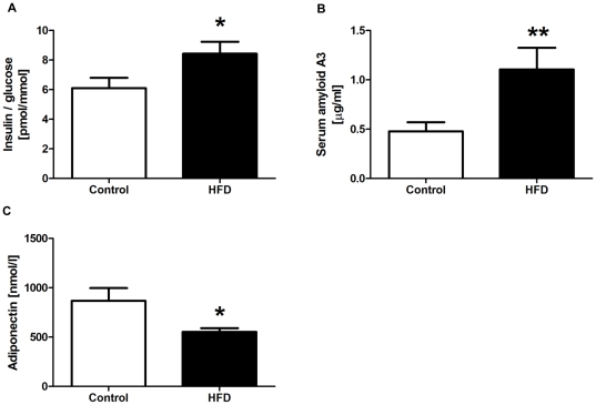
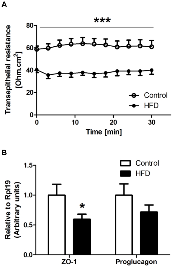
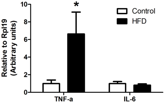
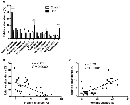
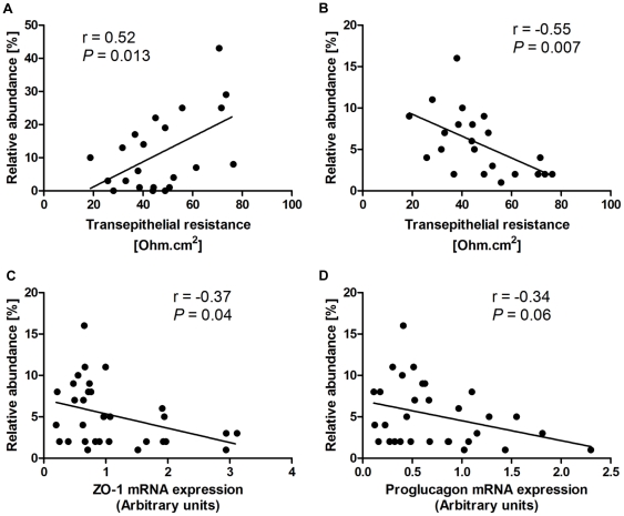
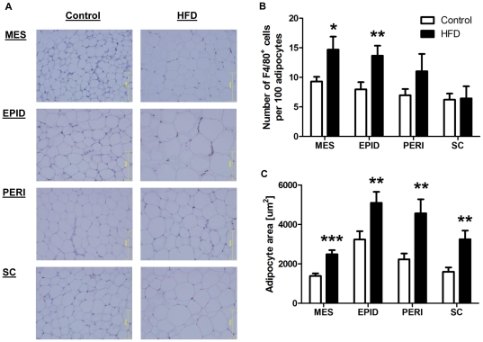
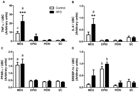
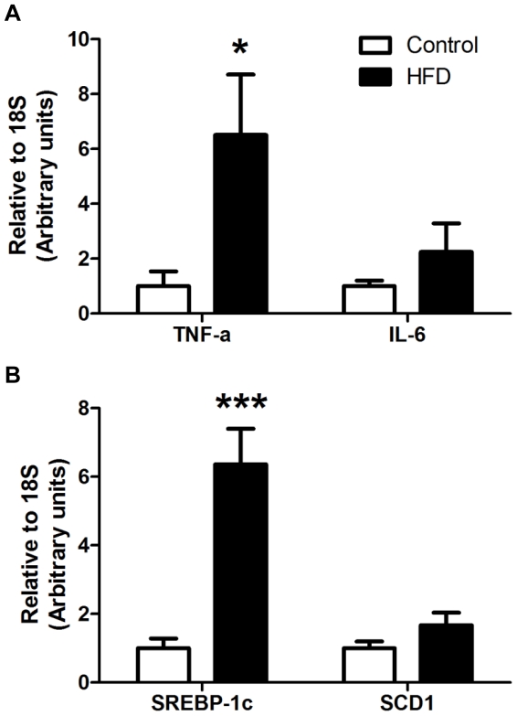
We investigated the relationship between gut health, visceral fat dysfunction and metabolic disorders in diet-induced obesity. C57BL/6J mice were fed control or high saturated fat diet (HFD). Circulating glucose, insulin and inflammatory markers were measured. Proximal colon barrier function was assessed by measuring transepithelial resistance and mRNA expression of tight-junction proteins. Gut microbiota profile was determined by 16S rDNA pyrosequencing. Tumor necrosis factor (TNF)-α and interleukin (IL)-6 mRNA levels were measured in proximal colon, adipose tissue and liver using RT-qPCR. Adipose macrophage infiltration (F4/80⁺) was assessed using immunohistochemical staining. HFD mice had a higher insulin/glucose ratio (P = 0.020) and serum levels of serum amyloid A3 (131%; P = 0.008) but reduced circulating adiponectin (64%; P = 0.011). In proximal colon of HFD mice compared to mice fed the control diet, transepithelial resistance and mRNA expression of zona occludens 1 were reduced by 38% (P<0.001) and 40% (P = 0.025) respectively and TNF-α mRNA level was 6.6-fold higher (P = 0.037). HFD reduced Lactobacillus (75%; P<0.001) but increased Oscillibacter (279%; P = 0.004) in fecal microbiota. Correlations were found between abundances of Lactobacillus (r = 0.52; P = 0.013) and Oscillibacter (r = -0.55; P = 0.007) with transepithelial resistance of the proximal colon. HFD increased macrophage infiltration (58%; P = 0.020), TNF-α (2.5-fold, P<0.001) and IL-6 mRNA levels (2.5-fold; P = 0.008) in mesenteric fat. Increased macrophage infiltration in epididymal fat was also observed with HFD feeding (71%; P = 0.006) but neither TNF-α nor IL-6 was altered. Perirenal and subcutaneous adipose tissue showed no signs of inflammation in HFD mice. The current results implicate gut dysfunction, and attendant inflammation of contiguous adipose, as salient features of the metabolic dysregulation of diet-induced obesity.


Conflict of interest statement

Competing Interests: I have read the journal's policy and have the following conflicts: JO and LHS are or have been employed by AstraZeneca. This does not alter our adherence to all the PLoS ONE policies on sharing data and materials. The other authors have declared that no competing interests exist.

Figures

Figure 1
Figure 1. Serum biochemical analyses.
Insulin/glucose ratio (A), concentrations of serum amyloid A3 (B) and adiponectin (C) in control and high saturated fat diet (HFD) fed mice (n = 15–16 per group). Data are shown as mean ± SEM. **P<0.01 and *P<0.05 compared to control.
Figure 2
Figure 2. Effect of high saturated fat diet (HFD) on barrier function of proximal colon.
A: Transepithelial resistance of mouse proximal colon was determined in the Ussing chamber by measuring the change in potential difference in response to 3 µA current generated across the tissue segment. B: mRNA levels of zona occludens (ZO)-1 and proglucagon were measured using RT-qPCR. Gene expression was normalized to RPL-19. Open circles/bars = control; Closed circles/bars = HFD (n = 9–16 per group). Data are shown as mean ± SEM. ***P<0.001 and *P<0.05 compared to control.
Figure 3
Figure 3. Effect of high saturated fat diet (HFD) on markers of inflammation in proximal colon.
mRNA expression of indicated genes was measured by RT-qPCR as described in Figure 2. Open bars = control; Closed bars = HFD (n = 11–16 per group). Data are shown as mean ± SEM. *P<0.05 compared to control.
Figure 4
Figure 4. Gut microbiota composition in control and high saturated fat diet (HFD) fed mice.
A: Relative abundance of main genera in fecal samples from control (open bars) and HFD (closed bars) mice (n = 16 per group). Data are shown as mean ± SEM. ***P<0.001, ** P<0.01 and *P<0.05 compared to control. Correlations between the abundance of Lactobacillus (B) and Oscillibacter (C) and weight changes. Gut microbiota profile was determined by metagenomic pyrosequencing from bacterial lineages in fecal samples.
Figure 5
Figure 5. Correlations between Lactobacillus (A) and Oscillibacter (B–D) abundances and permeability parameters in proximal colon.
Gut microbiota profile was determined as described in Figure 4 (n = 22–31). Transepithelial resistance and mRNA expression of zona occludens (ZO)-1 and proglucagon in proximal colon were measured as described in Figure 3.
Figure 6
Figure 6. Effect of high saturated fat diet (HFD) on adipose macrophage infiltration and adipocyte size.
A: Representative immunohistochemical staining of mesenteric (MES), epididymal (EPID), perirenal (PERI) and subcutaneous (SC) fat. Quantification of macrophage (F4/80+) infiltration (B) and adipocyte size (C). Tissues were fixed in formalin and embedded in paraffin. Sections were stained for F4/80 and counterstained with hematoxylin. Open bars = control; Closed bars = HFD (n = 4–10 per group). Data are shown as mean ± SEM. ***P<0.001, **P<0.01 and *P<0.05 compared to control.
Figure 7
Figure 7. mRNA expression of TNF-α (A), IL-6 (B), PPAR-γ (C) and SREBP-1c (D) in adipose tissue.
mRNA levels in control and high saturated fat diet (HFD) fed mice were measured by RT-qPCR. Gene expression was normalized to UBC. Open bars = control; Closed bars = HFD (n = 12–15 per group). Data are shown as mean ± SEM. ***P<0.001 and **P<0.01 compared to control. a, b, c: P<0.001, P<0.01 and P<0.05 respectively compared to other depots in the same diet group.
Figure 8
Figure 8. mRNA expression of genes involved in inflammation (A) and lipogenesis (B) in liver.
mRNA levels of TNF-α, IL-6, SREBP-1c and SCD1 in control and high saturated fat diet (HFD) fed mice were measured using RT-qPCR. Gene expression was normalized to 18S. Open bars = control; Closed bars = HFD (n = 14–16 per group). Data are shown as mean ± SEM. ***P<0.001 and *P<0.05 compared to control.

References

    1. Miyazaki Y, Glass L, Triplitt C, Wajcberg E, Mandarino LJ, et al. Abdominal fat distribution and peripheral and hepatic insulin resistance in type 2 diabetes mellitus. Am J Physiol Endocrinol Metab. 2002;283:E1135–1143. - PubMed
    1. Sam S, Haffner S, Davidson MH, D'Agostino RB, Sr, Feinstein S, et al. Relation of abdominal fat depots to systemic markers of inflammation in type 2 diabetes. Diabetes Care. 2009;32:932–937. - PMC - PubMed
    1. Barbarroja N, Lopez-Pedrera R, Mayas MD, Garcia-Fuentes E, Garrido-Sanchez L, et al. The obese healthy paradox: is inflammation the answer? Biochem J. 2010;430:141–149. - PubMed
    1. Lam YY, Janovska A, McAinch AJ, Belobrajdic DP, Hatzinikolas G, et al. The use of adipose tissue-conditioned media to demonstrate the differential effects of fat depots on insulin-stimulated glucose uptake in a skeletal muscle cell line. Obes Res Clin Pract. 2011;5:e43–e54. - PubMed
    1. Fain JN, Madan AK, Hiler ML, Cheema P, Bahouth SW. Comparison of the release of adipokines by adipose tissue, adipose tissue matrix, and adipocytes from visceral and subcutaneous abdominal adipose tissues of obese humans. Endocrinology. 2004;145:2273–2282. - PubMed

Publication types