. 2012 Mar 29:9:61.
doi: 10.1186/1742-2094-9-61.

Peroxisome deficiency but not the defect in ether lipid synthesis causes activation of the innate immune system and axonal loss in the central nervous system

Affiliations

Peroxisome deficiency but not the defect in ether lipid synthesis causes activation of the innate immune system and axonal loss in the central nervous system

Astrid Bottelbergs et al. J Neuroinflammation. .

Abstract

Background: Mice with peroxisome deficiency in neural cells (Nestin-Pex5-/-) develop a neurodegenerative phenotype leading to motor and cognitive disabilities and early death. Major pathologies at the end stage of disease include severe demyelination, axonal degeneration and neuroinflammation. We now investigated the onset and progression of these pathological processes, and their potential interrelationship. In addition, the putative role of oxidative stress, the impact of plasmalogen depletion on the neurodegenerative phenotype, and the consequences of peroxisome elimination in the postnatal period were studied.

Methods: Immunohistochemistry in association with gene expression analysis was performed on Nestin-Pex5-/- mice to document demyelination, axonal damage and neuroinflammation. Also Gnpat-/- mice, with selective plasmalogen deficiency and CMV-Tx-Pex5-/- mice, with tamoxifen induced generalized loss of peroxisomes were analysed.

Results: Activation of the innate immune system is a very early event in the pathological process in Nestin-Pex5-/- mice which evolves in chronic neuroinflammation. The complement factor C1q, one of the earliest up regulated transcripts, was expressed on neurons and oligodendrocytes but not on microglia. Transcripts of other pro- and anti-inflammatory genes and markers of phagocytotic activity were already significantly induced before detecting pathologies with immunofluorescent staining. Demyelination, macrophage activity and axonal loss co-occurred throughout the brain. As in patients with mild peroxisome biogenesis disorders who develop regressive changes, demyelination in cerebellum and brain stem preceded major myelin loss in corpus callosum of both Nestin-Pex5-/- and CMV-Tx-Pex5-/- mice. These lesions were not accompanied by generalized oxidative stress throughout the brain. Although Gnpat-/- mice displayed dysmyelination and Purkinje cell axon damage in cerebellum, confirming previous observations, no signs of inflammation or demyelination aggravating with age were observed.

Conclusions: Peroxisome inactivity triggers a fast neuroinflammatory reaction, which is not solely due to the depletion of plasmalogens. In association with myelin abnormalities this causes axon damage and loss.


Figures
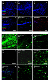
Figure 1
Figure 1
Early myelin abnormalities but mild microgliosis in cerebellum. (A-F) Immunohistochemistry was performed to visualize microglia (F4/80, green), damaged axons (SMI32, red) and myelin (MBP, blue). Compared to the control mice (A) several fibers in the cerebellar folia of the Nestin-Pex5−/− already lack myelin at two weeks (B), which was even more pronounced at three weeks (C-D) and six weeks (E-F). SMI32 positive axons, indicative of degeneration, were observed at six weeks (F). (G-I) Axonal loss was analyzed by the use of an antibody recognizing healthy phosphorylated axons (SMI31). Decreased SMI31 immunoreactivity was observed at six (H) and 12 weeks (I), in comparison with the control littermates (G). In addition, several axonal swellings were present (H-I, arrows). (J-L) Astrocytes were visualized by anti-GFAP, which revealed higher immunoreactivity at three weeks in white matter of the folia, but also in the molecular layer (K). At 12 weeks astrocyte proliferation and activation was even more pronounced (L). (M) Double labeling of myelin (MBP, blue) and activated/phagocytotic microglia with MAC-3 (green) in the cerebellum of a nine-week-old Nestin-Pex5−/− mouse. (N-O) An antibody specific for potassium channels (K+) was used for the investigation of paranodal structures. Cerebellar axons displayed an abnormal distribution of potassium channels (O, arrows), as compared to the strict juxtaparanodal localization in the control (N). All panels of each row were stained with the same antibodies. Scale bars: A-M: 100 μm; N-O: 10 μm. GFAP: glial fibrillary acidic protein, K+: potassium, MAC: macrophage, MBP: myelin basic protein, SMI: Sternberger Monoclonals Inc.
Figure 2
Figure 2
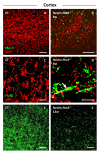
Myelin loss and mild microgliosis in the cortex. (A-D) Double immunolabeling of myelin (MBP, red) and activated microglia (MAC-3, green) or microglia (F4/80, green), respectively. Cortical demyelination was detected in a subset of Nestin-Pex5−/− mice at six weeks (B and D), but a strong microglial reaction was never seen (B and D). However, the observed microglial cells were MAC-3 positive, which indicates phagocytotic activity (B) and some microglia seem to contain myelin (D, arrow). (E-F) Loss of axons, represented as a decreased SMI31 immunoreactivity, was clearly detectable in the cortex of 12-week-old knockout mice (F), when compared to the control mice (E). All panels of each row were stained with the same antibodies. Scale bars: A-C and G-I: 100 μm; D-F: 25 μm. MBP, myelin basic protein.
Figure 3
Figure 3
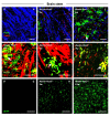
Association of severe demyelination with extensive microgliosis in the brain stem. (A-C) Triple staining of microglia (F4/80, green), damaged axons (SMI32, red) and myelin (MBP, blue). Already at the age of three and six weeks demyelinated and degenerating axons are observed in a subset of Nestin-Pex5−/− mice (B-C), in contrast with the control littermates (A). Microglial cells are observed in association with these demyelinated axons (C, arrow). (D-F) Higher magnification of the double labeling of myelin (MBP, red) and microglia (F4/80, green) in the brain stem of six- to nine-week-old Nestin-Pex5−/− mice. The observed microglial cells have a swollen appearance and several of them contain MBP-positive myelin debris (D-F, arrows). (G-I) The anti-GFAP antibody was used to analyze astrogliosis, which is mild at three weeks (H), but strongly pronounced at 12 weeks (I), compared with the control mice (G). All panels of each row were stained with the same antibodies. Scale bars: A-D and G-I: 100 μm; E-F: 50 μm. GFAP, glial fibrillary acid protein; MBP, myelin basic protein.
Figure 4
Figure 4
Corpus callosum is affected later than other brain regions. (A-B) Microglia (F4/80, green), axonal swellings (APP, red) or damage (SMI32, red) and myelin (MBP, blue) were co-labeled on sagittal brain section. At six weeks no pathology was observed yet in the corpus callosum, but at 12 weeks demyelination was pronounced and associated with microgliosis (B) and axonal swellings (Inset in B, arrow). (C-D) Healthy axons were massively lost at 12 weeks, as shown by decreased SMI31 immunoreactivity (D), compared to control mice (C). (E-F) Potassium channels (K+) were immunolabeled to visualize the juxtaparanodes. In the 12-week-old Nestin-Pex5−/− mice these juxtaparanodes displayed a broader distribution (F, arrow), in comparison with the control mice (E). (G-H) Astrocytes were labeled with anti-GFAP. Astrogliosis is strongly pronounced at 12 weeks (H), in comparison with the control mouse (G). Scale bars: A-D: 100 μm; G-H: 10 μm; Inset in B: 12 μm. APP: amyloid precursor protein, GFAP: glial fibrillary acidic protein, K+: potassium, MBP: myelin basic protein, SMI: Sternberger monoclonals Inc.
Figure 5
Figure 5
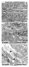
Ultrastructure of myelin and axons in corpus callosum. (A) Cross and longitudinal sections of nerve fibers of control mice display a regular and compact myelin sheath. (B) In the corpus callosum of three-week-old Nestin-Pex5−/− mice many axons are still myelinated while a subset already lacks a myelin sheath. The arrow in (B) indicates a swollen necrotic axon, surrounded by a normally appearing myelin sheath. (C) In 12-week-old Nestin-Pex5 knockout mice demyelination is even more pronounced than at three weeks. Similarly as at three weeks, an edematous necrotic axon, which is enwrapped by an apparently normal myelin sheath (arrow), is detected. In addition, large necrotic areas are observed (asterisk).
Figure 6
Figure 6
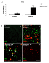
C1qexpression is an early response in Nestin-Pex5−/− brain. (A) C1q expression levels were quantified by qRT-PCR. Up regulation of mRNA levels was already significant in the corpus callosum of the three-week-old Nestin-Pex5−/−, although this was much more pronounced at six weeks. Expression levels were normalized to β-actin expression. Values are given as means ± SEM of four independent samples. ** P < 0.01, *** P < 0.001 (Student’s t-test). (B-E) C1q was also investigated by immunohistochemistry. At six weeks, C1q immunoreactivity was observed in the corpus callosum (D) and brain stem (C), but not in the cerebellum (E). Double labeling of C1q (green) and microglia (F4/80, red) or oligodendrocytes (CC-1, red), respectively, was performed to determine the cellular localization of the complement protein. C1q was not present in microglia (B and D), but in a subset of oligodendrocytes (C). Scale bars: B and E: 50 μm; C and D: 20 μm. SEM, standard error of the mean.
Figure 7
Figure 7
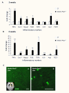
Up regulation of pro- and anti-inflammatory markers in Nestin-Pex5−/− mice. (A-B) In the corpus callosum of three- and six-week-old Nestin-Pex5−/− mice mRNA levels of several pro- and anti-inflammatory markers were quantified by qRT-PCR. (A) TNFα***, Cxcl-1*, Mpeg1** and TLR2* were all significantly up regulated in the corpus callosum of three-week-old knockout mice. The anti-inflammatory markers TGFβ, IL10, arginase 1 and FIZZ1 were not up regulated at three weeks. (B) At six weeks the pro-inflammatory markers showed much higher expression levels, compared to three-week-old Nestin-Pex5 knockout mice. Also expression levels of the anti-inflammatory markers TGFβ, IL10 and arginase 1 were significantly increased at this age. Values represent means ± SEM of four independent samples, * P < 0.05, ** P < 0.01, *** P < 0.001 (Student’s t-test). Expression levels were normalized to β-actin expression. (C-D) By immunohistochemistry infiltration of CD3 positive lymphocytes was observed in 12-week-old Nestin-Pex5−/−mice, mainly in the corpus callosum. Scale bars: C: 50 μm; D: 25 μm. CD: cluster of differentiation, Cxcl: chemokine (C-X-C motif) ligand, FIZZ: found in inflammatory zone, IL: interleukin, Mpeg: macrophage expressed gene, SEM: standard error of the mean, TGF: transforming growth factor, TLR: toll like receptor.
Figure 8
Figure 8
Oxidative stress markers are only present in Purkinje cells. (A) End products of lipid peroxidation, predominantly malondialdehyde, were quantified by the TBARS assay. No differences between 9- to 12-week-old Nestin-Pex5−/− and control littermates were observed in the corpus callosum, cerebellum or spinal cord. Values are given as means ± SEM of four independent samples of each genotype (Student’s t-test). (B-I) The markers 4-HNE (B-E) and 3-nitrotyrosine (3-NT) (F-I) were evaluated by immunohistochemistry. 4-HNE was slightly observable in Purkinje cells of two-week-old Nestin-Pex5−/− mice (C), compared with the control littermates (B). The amount of 4-HNE present in Purkinje cells was higher at three (D) and 12 weeks (E). Low 3-NT immunoreactivity was observed at three weeks in Purkinje cells (G), but increased with age (H-I). Scale bars: B-F and G: 100 μm; G and I: 50 μm. 4-HNE, 4-hydroxynonenal; SEM, standard error of the mean; TBARS, thiobarbituric acid reactive substances.
Figure 9
Figure 9
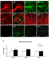
Cerebellar and cortical dysmyelination inGnpat−/− mice in the absence of microglia activation. (A-L) Immunohistochemistry was performed to investigate myelin (MBP), axonal loss (SMI31), microgliosis (F4/80) and oxidative stress (4-HNE and 3-nitrotyrosine). Gnpat−/− mice displayed a decreased amount of myelin in the cerebellar folia at three months (B), compared with the control littermates (A). Cortical demyelination was observed in a subset of three-month-old Gnpat knockout mice (G-H). Axonal loss was very mild at three weeks (C-D) and five months (E-F) and also a few axonal swellings were detectable with SMI31 (red) at five months (F, arrow and inset). Microgliosis (F4/80, green) was absent (F). 3-Nitrotyrosine (3-NT) and 4-HNE positive Purkinje cells were seen in three-week-old Gnpat−/− mice (J, L), in comparison with the control mice (I, K). Scale bars: A-F and I-L: 100 μm; G-H: 50 μm. (M) Quantification of mRNA levels of the pro-inflammatory markers TNFα, TLR2 and C1q by qRT-PCR. Expression levels were not elevated in four- to five-month-old Gnpat knockout mice. Values represent means ± SEM of four independent samples. Expression levels were normalized to β-actin expression. 4-HNE, 4-hydroxynonenal; TLR2, toll-like receptor 2; SEM, standard error of the mean.

Similar articles

Cited by

References

    1. Steinberg S, Dodt G, Raymond GV, Braverman NE, Moser AB, Moser HW. Peroxisome biogenesis disorders. Biochim Biophys Acta. 2006;1763:1733–1748. doi: 10.1016/j.bbamcr.2006.09.010. - DOI - PubMed
    1. Wanders RJ, Waterham HR. Peroxisomal disorders: the single peroxisomal enzyme deficiencies. Biochim Biophys Acta. 2006;1763:1707–1720. doi: 10.1016/j.bbamcr.2006.08.010. - DOI - PubMed
    1. Baes M, Aubourg P. Peroxisomes, myelination, and axonal integrity in the CNS. Neuroscientist. 2009;15:367–379. doi: 10.1177/1073858409336297. - DOI - PubMed
    1. Hulshagen L, Krysko O, Huyghe S, Klein R, Van Veldhoven PP, De Deyn PP, D’hooge R, Hartmann D, Baes M. Absence of functional peroxisomes from mouse central nervous system causes dysmyelination and axon degeneration. J Neurosci. 2008;28:4015–4027. doi: 10.1523/JNEUROSCI.4968-07.2008. - DOI - PMC - PubMed
    1. Krysko O, Hulshagen L, Janssen A, Schutz G, Klein R, De Bruycker M, Espeel M, Gressens P, Baes M. Neocortical and cerebellar developmental abnormalities in conditions of selective elimination of peroxisomes from brain or from liver. J Neurosci Res. 2007;85:58–72. doi: 10.1002/jnr.21097. - DOI - PubMed

Publication types

MeSH terms