. 2012 Mar 29:9:27.
doi: 10.1186/1742-4690-9-27.

Tailored enrichment strategy detects low abundant small noncoding RNAs in HIV-1 infected cells

Affiliations

Tailored enrichment strategy detects low abundant small noncoding RNAs in HIV-1 infected cells

Claudia F Althaus et al. Retrovirology. .

Abstract

Background: The various classes of small noncoding RNAs (sncRNAs) are important regulators of gene expression across divergent types of organisms. While a rapidly increasing number of sncRNAs has been identified over recent years, the isolation of sncRNAs of low abundance remains challenging. Virally encoded sncRNAs, particularly those of RNA viruses, can be expressed at very low levels. This is best illustrated by HIV-1 where virus encoded sncRNAs represent approximately 0.1-1.0% of all sncRNAs in HIV-1 infected cells or were found to be undetected. Thus, we applied a novel, sequence targeted enrichment strategy to capture HIV-1 derived sncRNAs in HIV-1 infected primary CD4+ T-lymphocytes and macrophages that allows a greater than 100-fold enrichment of low abundant sncRNAs.

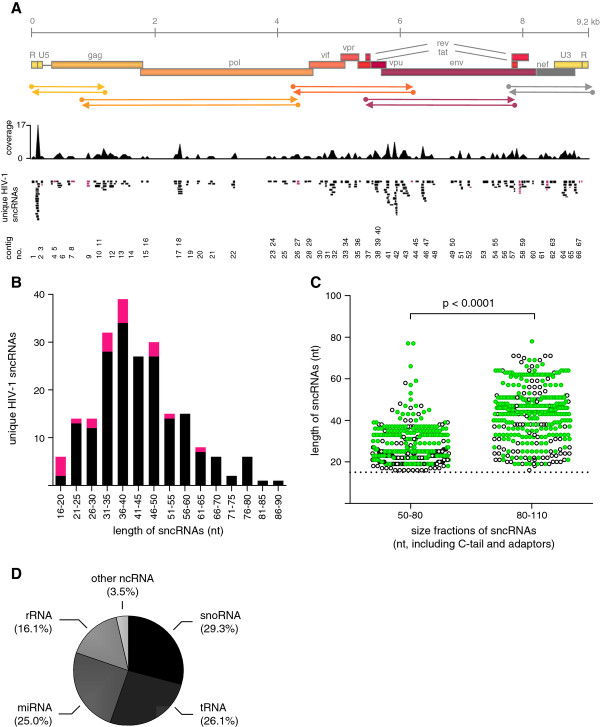
Results: Eight hundred and ninety-two individual HIV-1 sncRNAs were cloned and sequenced from nine different sncRNA libraries derived from five independent experiments. These clones represent up to 90% of all sncRNA clones in the generated libraries. Two hundred and sixteen HIV-1 sncRNAs were distinguishable as unique clones. They are spread throughout the HIV-1 genome, however, forming certain clusters, and almost 10% show an antisense orientation. The length of HIV-1 sncRNAs varies between 16 and 89 nucleotides with an unexpected peak at 31 to 50 nucleotides, thus, longer than cellular microRNAs or short-interfering RNAs (siRNAs). Exemplary HIV-1 sncRNAs were also generated in cells infected with different primary HIV-1 isolates and can inhibit HIV-1 replication.

Conclusions: HIV-1 infected cells generate virally encoded sncRNAs, which might play a role in the HIV-1 life cycle. Furthermore, the enormous capacity to enrich low abundance sncRNAs in a sequence specific manner highly recommends our selection strategy for any type of investigation where origin or target sequences of the sought-after sncRNAs are known.


Figures

Figure 1
Figure 1
Strategy of cDNA library generation with hybridization capture for HIV-1 encoded small noncoding RNAs (sncRNAs). HIV-1 susceptible cells (in our set-up, primary human macrophages or CD4+ T-lymphocytes) are infected with HIV-1 (Step 1). Cellular (black) and HIV-1 encoded (bright green) sncRNAs (< 200 nt) are extracted from HIV-1 infected cells (Step 2). RNA is C-tailed at the 3'-end, adaptor-ligated at the 5'-end (Step 3), and RT-PCR is performed (Step 4). For the preparation of the HIV-1 ssDNA hybridization probes, PCR is performed with biotinylated primers for 5 overlapping regions of the genome using HIV-1JR-FL plasmid as template. Biotinylated amplicons are attached to streptavidin beads (Box 1). Sequences homologous to HIV-1 are enriched by incubation of cDNA derived from adaptor-ligated sncRNAs with a mixture of the 5 different HIV-1 ssDNA hybridization probes (Step 5); alternatively each HIV-1 ssDNA hybridization probe can be used separately. After hybridization capture, bound amplicons are eluted, amplified, and size selected on a gel (Step 6). The hybridization and size selection steps can be repeated (Step 7). Amplicons are cloned and sequenced or can be sequenced using next-generation sequencing technologies (Step 8).
Figure 2
Figure 2
Efficiency of hybridization capture to enrich HIV-1 encoded sncRNAs. Comparison of published data using the currents standard protocols (left) with our novel selection strategy (right). Standard protocols with no selection led to a yield of 0.1-1.0% HIV-1 sncRNAs. Numbers above bars indicate the respective literature references [8,11,13]. Using our novel method, application of one round of hybridization capture yielded 8.9 ± 5.7% (mean ± SD) HIV-1 sncRNAs (libraries A and B). Performing two consecutive rounds of selection (libraries C-J) optimized the yield to 78.3 ± 7.6% (mean ± SD) HIV-1 sncRNAs.
Figure 3
Figure 3
Alignment and characterization of identified sncRNAs. (A) 216 unique sncRNAs from nine libraries (Supplementary dataset 1) were aligned to the reference strain HIV-1HXB2 and cluster in 67 contigs distributed throughout the whole genome of HIV-1. The coverage of sncRNA per nucleotide, the single unique clones (black: sense HIV-1 sncRNAs, pink: antisense HIV-1 sncRNAs), and the contig numbers are shown. (B) The length (nucleotides, nt) distribution of all unique HIV-1 sncRNAs is depicted. Sense sncRNAs are shown in black, antisense sncRNAs are shown in pink. (C) Probing the influence of target molecule length on hybridization efficacy, libraries F, G, H, and J underwent a second size separation step before undergoing a second round of hybridization enrichment. Dehybridized cDNA was separated into two fractions of 80-110 bp or 50-80 bp length and both probed separately for hybridization efficacy. Green full circles denote HIV-1 derived sncRNAs, black open circles denote non-HIV-1 sncRNAs. Both fractions successfully retrieved sncRNA in the second round hybridization. 20-25 nucleotide long sncRNAs were retrieved from both fractions and comprised 40.1% of all sncRNAs in the small size fraction and 11.1% in the large size fraction (p < 0.0001, Chi square test). (D) Pie chart depicting the distribution of different human cellular sncRNAs in all libraries.
Figure 4
Figure 4
Functional analysis of HIV-1 sncRNAs. (A) CD4+ T-lymphocytes of three healthy donors were infected with the indicated five different HIV-1 primary isolates and screened for the presence of HIV-1 sncRNAs in 3 contigs (contigs 2, 43, and 58) identified in HIV-1JR-FL infected cultures. Cell cultures from three HIV-1 negative donors were probed for each virus, and the cultures which scored positive for the respective HIV-1 sncRNAs are depicted in red squares. (B) Primary macrophages and CD8+ T-cell depleted PBMCs of two healthy donors were infected with HIV-1JR-FL and HIV-1 sncRNA contig 2 (green) and contig 58 (orange) which were quantified by qPCR 14 (macrophages) and 6 (CD8+ T-cell depleted PBMC) days post infection. Amplification was unsuccessful in non-infected cells (data not depicted). As controls, the cellular miRNAs hsa-miR-21 (white and light grey for non-infected cells) and hsa-miR-223 (black and dark grey for non-infected cells) were quantified. < d.l., below detection limit of 1 RNA copy/103 cells. (C) Predicted secondary structures of sncRNALTR6, hybrids of sense and antisense orientated clones from contig 58, namely sncRNAenv183, sncRNAenv184 and sncRNAenv185, and the positive control siRNA-M184 pol [24] (Additional file 3: Table S3) are depicted. (D) Inhibition of HIV-1 replication in primary macrophages by HIV-1 sncRNAs. Macrophages were infected with HIV-1JR-FL seven days before transfection with sncRNAs. On day 0 cells were transfected with 50 nM of the indicated sncRNA (sncRNALTR6 (green), sncRNAenv183&185 (orange), sncRNAenv184&185 (red)) and viral replication was monitored by p24 ELISA on days 0, 5, 9, and 13 post transfection. Mock transfected (black), scrambled sncRNA (blue) and siRNA nonsense (grey) were used as negative control, and siRNA-M184 pol [20] (open circles) as positive control. (E) Western blot of primary macrophage lysates probed with anti-MxA antibody or anti-β-actin antibody as control. Primary macrophages were transfected with siRNA nonsense, sncRNAenv183&185, and sncRNAenv184&185 or treated with interferon-αA/D (positive control). The negative control comprised of untreated and untransfected macrophages.

References

    1. Fire A, Xu S, Montgomery MK, Kostas SA, Driver SE, Mello CC. Potent and specific genetic interference by double-stranded RNA in Caenorhabditis elegans. Nature. 1998;391:806–811. doi: 10.1038/35888. - DOI - PubMed
    1. Farazi TA, Juranek SA, Tuschl T. The growing catalog of small RNAs and their association with distinct Argonaute/Piwi family members. Development. 2008;135:1201–1214. doi: 10.1242/dev.005629. - DOI - PubMed
    1. Filipowicz W, Bhattacharyya SN, Sonenberg N. Mechanisms of post-transcriptional regulation by microRNAs: are the answers in sight? Nat Rev Genet. 2008;9:102–114. - PubMed
    1. Huttenhofer A, Vogel J. Experimental approaches to identify non-coding RNAs. Nucleic Acids Res. 2006;34:635–646. doi: 10.1093/nar/gkj469. - DOI - PMC - PubMed
    1. Kong W, Zhao JJ, He L, Cheng JQ. Strategies for profiling microRNA expression. J Cell Physiol. 2009;218:22–25. doi: 10.1002/jcp.21577. - DOI - PubMed

Publication types