. 2012 Jun;220(6):591-602.
doi: 10.1111/j.1469-7580.2012.01497.x. Epub 2012 Mar 28.

The generation of vertebral segmental patterning in the chick embryo

Affiliations

The generation of vertebral segmental patterning in the chick embryo

Biruntha Senthinathan et al. J Anat. 2012 Jun.

Abstract

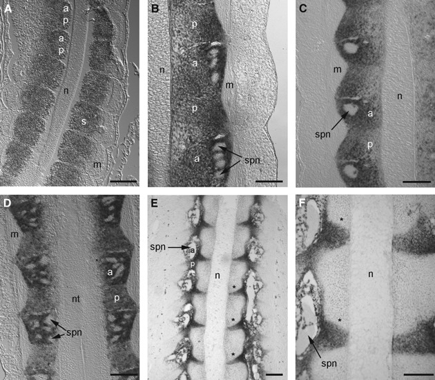
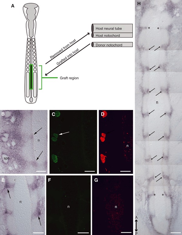
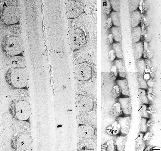
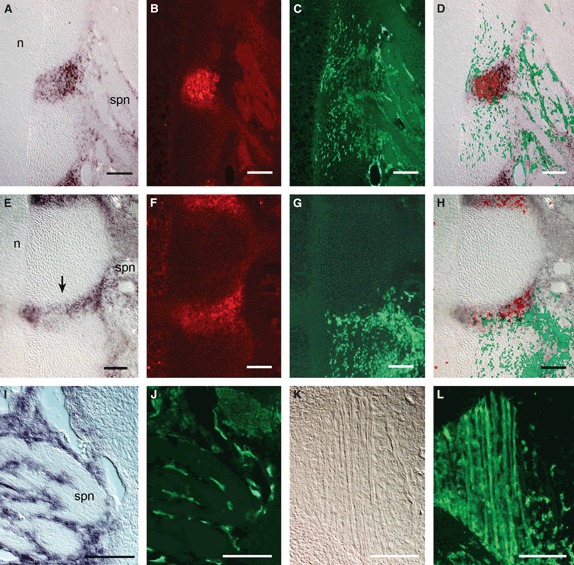
We have carried out a series of experimental manipulations in the chick embryo to assess whether the notochord, neural tube and spinal nerves influence segmental patterning of the vertebral column. Using Pax1 expression in the somite-derived sclerotomes as a marker for segmentation of the developing intervertebral disc, our results exclude such an influence. In contrast to certain teleost species, where the notochord has been shown to generate segmentation of the vertebral bodies (chordacentra), these experiments indicate that segmental patterning of the avian vertebral column arises autonomously in the somite mesoderm. We suggest that in amniotes, the subdivision of each sclerotome into non-miscible anterior and posterior halves plays a critical role in establishing vertebral segmentation, and in maintaining left/right alignment of the developing vertebral elements at the body midline.


Figures

Fig. 1
Fig. 1
Pax1 expression in the developing chick sclerotome. Longitudinal sections, anterior upwards. (A) Stage 13 embryo at the level of the notochord, showing uniform Pax1 expression in both anterior (a) and posterior (p) halves of each sclerotome. (B) Stage 24 embryo, thoracic somites; uniform Pax1 expression persists in both sclerotome halves. (C, D) Stage 25 embryo, thoracic somites, at the level of the notochord (C) and neural tube (D), showing more intense Pax1 expression in the anterior half-sclerotomes surrounding the spinal nerves. (E) Stage 29 embryo and (F) stage 30 embryo, showing resolution of Pax1 expression into segmentally repeated stripes of cells (asterisks) extending medially from the spinal nerves and converging on the notochord. a, anterior half-sclerotome; m, myotome; n, notochord; nt, neural tube; p, posterior half-sclerotome; s, sclerotome; spn, spinal nerve. Scale bars: 100 μm.
Fig. 2
Fig. 2
Notochord excision. (A) Schematic diagram showing experimental strategy for notochordectomy. The dashed lines (a’ and a’’) show the levels of the sections shown in (Aa’) and (Aa’’). (Aa’) Transverse section through the unoperated region of an embryo fixed immediately after excision of the neural tube and notochord, and processed for shh in situ hybridization; shh expression (red) is visible in the notochord. (Aa’’) Transverse section of the same embryo through the excised region and processed for shh in situ hybridization; removal of the notochord and its sheath is confirmed, and the midline endoderm remains intact. (B) Longitudinal section of a stage 29 host embryo showing the unoperated region with notochord (posterior, downwards) and the notochordectomized region (anterior, upwards). Pax1 expression remains segmented in the grafted region anterior to the notochord (arrows), being conspicuous laterally but diminished medially in comparison with the stripes of expression seen alongside the notochord in the unoperated region. (C) Similar section to (B) in another notochordectomized embryo, showing persistent Pax1 expression both laterally and medially (arrows) in the grafted region anterior to the notochord. n, notochord. Scale bars: 100 μm.
Fig. 3
Fig. 3
Neural tube excision. (A) Schematic diagram showing experimental strategy for neuralectomy. (B–D) Longitudinal sections (anterior upwards) of the unoperated region of a stage 29 host embryo. Pax1 expression is shown in a bright-field image (B), present in two horizontal stripes (arrows) adjacent to the notochord, and in the vicinity of the spinal nerves. Spinal nerves also stain for TUJ1 (C; arrow) and HNK1 (D) expression, and some notochord cells also stain weakly for HNK1. (E–G) Longitudinal sections (anterior upwards) of neuralectomized region at the level of the notochord, showing stripes of Pax1 expression (arrows) adjacent to the notochord (E), in the absence of TUJ1 and HNK1 expression (F, G) confirming removal of spinal nerve components; weak HNK1 expression persists in notochord cells. (H) Composite longitudinal section at the level of the notochord along the entire length of the operated region of a neuralectomized embryo, anterior (A) and posterior (P) directions as indicated. Pax1 expression (arrows) remains segmented at the level of the notochord. Stripes were occasionally somewhat thicker along the anterior–posterior axis than in normal embryos, perhaps indicating that the neural tube and/or spinal nerves influence overall growth of the annulus fibrosus. The segments marked with an asterisk lie at the boundaries of the operated and unoperated regions. n, notochord; spn, spinal nerve. Scale bars: 100 μm.
Fig. 4
Fig. 4
Reversal of PSM unilaterally along the anterior–posterior axis. (A) Longitudinal section through stage 29 host embryo at the level of the neural arches and neural tube, anterior upwards. Numbers indicate individual dorsal root ganglia, and the intervening neural arches (pedicles) are placed between them. Ganglia 3, 4 and 5 show left/right misalignment resulting from three-segment PSM reversal on the right-hand side. (B) Longitudinal section through a stage 29 host embryo at the level of the notochord, showing left/right misalignment of the stripes of Pax1 expression (arrows) in the region of PSM reversal. The characteristic curvature of the stripes is also inverted in the reversed region compared with the four normal segments placed more anteriorly on the right-hand side and with those on the left-hand side. Size differences between normal and reversed regions (e.g. for ganglia, neural arches and distances between adjacent Pax1 stripes) may reflect overall growth differences between these regions in the graft vs. host. n, notochord; na, neural arch; nt, neural tube; spn, spinal nerve. Scale bars: 100 μm.
Fig. 5
Fig. 5
Ectopic grafts of GFP-labelled somitocoele cells and epithelial somite cells. (A–D) Stage 31 embryo, longitudinal section after an ectopic graft of somitocoele cells between epithelial somite III and the neural tube/notochord. (A) Typical striped Pax1 expression in the graft region between the notochord and spinal nerves, as in normal embryos; (B) expression of calretinin in the Pax1-expressing stripe in the same section. (C) The wide distribution of grafted GFP-labelled cells in this section; (D) an overlay of (A–C). Despite extensive dispersal of grafted somitocoele-derived cells, Pax1 and calretinin expression show normal localization with no evidence of ectopic expression. (E, F) Stage 29 embryo, longitudinal section after an ectopic graft of somite epithelial cells between epithelial somite III and the neural tube/notochord. (E) Typical striped Pax1 expression in the graft region between adjacent vertebral bodies; (F) calretinin expression in the same stripe of cells. (G) Extensive dispersal of grafted GFP-labelled cells in this section; (H) the overlay of (E–G). Grafted epithelial cells are dispersed widely in both vertebral and intervertebral positions. (I–L) Stage 31 embryo, longitudinal sections after an ectopic graft of somitocoele cells between epithelial somite III and the neural tube/notochord. Pax1-expressing cells (I) and GFP-labelled cells (J) occupy positions adjacent to spinal axons; GFP-labelled cells also contribute to the myotome (K, bright-field; L, GFP expression). spn, spinal nerve. Scale bars: 100 μm.

Similar articles

Cited by

References

    1. Aoyama H, Asamoto K. The developmental fate of the rostral/caudal half of a somite for vertebra and rib formation: experimental confirmation of the resegmentation theory using chick-quail chimeras. Mech Dev. 2000;99:71–82. - PubMed
    1. Aszodi A, Chan D, Hunziker E, et al. Collagen II is essential for the removal of the notochord and the formation of intervertebral discs. J Cell Biol. 1998;143:1399–1412. - PMC - PubMed
    1. Baffi MO, Moran MA, Serra R. Tgfbr2 regulates the maintenance of boundaries in the axial skeleton. Dev Biol. 2006;296:363–374. - PMC - PubMed
    1. Bagnall KM, Sanders EJ. The binding pattern of peanut lectin associated with sclerotome migration and the formation of the vertebral axis in the chick embryo. Anat Embryol (Berl) 1989;180:505–513. - PubMed
    1. Bagnall KM, Higgins SJ, Sanders EJ. The contribution made by a single somite to the vertebral column: experimental evidence in support of resegmentation using the chick-quail chimaera model. Development. 1988;103:69–85. - PubMed

Publication types

Substances

LinkOut - more resources