Review
. 2012 Jul;52(1):44-51.
doi: 10.1016/j.ceca.2012.03.001. Epub 2012 Mar 28.

Mitochondrial Ca(2+) signals in autophagy

Affiliations
Review

Mitochondrial Ca(2+) signals in autophagy

César Cárdenas et al. Cell Calcium. 2012 Jul.

Abstract

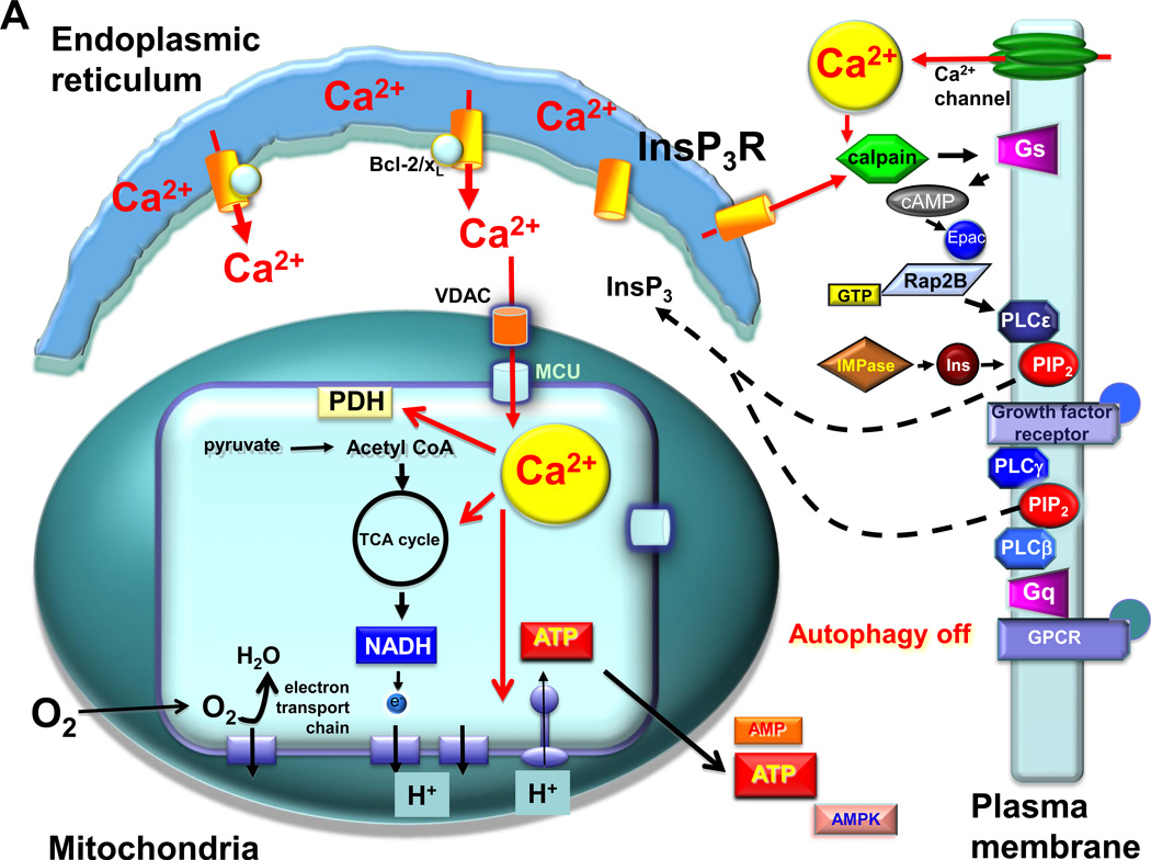
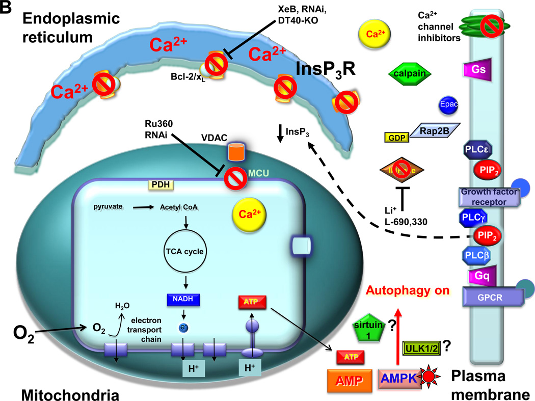
Macroautophagy (autophagy) is a lysosomal degradation pathway that is conserved from yeast to humans that plays an important role in recycling cellular constituents in all cells. A number of protein complexes and signaling pathways impinge on the regulation of autophagy, with the mammalian target of rapamycin (mTOR) as the central player in the canonical pathway. Cytoplasmic Ca(2+) signaling also regulates autophagy, with both activating and inhibitory effects, mediated by the canonical as well as non-canonical pathways. Here we review this regulation, with a focus on the role of an mTOR-independent pathway that involves the inositol trisphosphate receptor (InsP(3)R) Ca(2+) release channel and Ca(2+) signaling to mitochondria. Constitutive InsP(3)R Ca(2+) transfer to mitochondria is required for autophagy suppression in cells in nutrient-replete media. In its absence, cells become metabolically compromised due to insufficient production of reducing equivalents to support oxidative phosphorylation. Absence of this Ca(2+) transfer to mitochondria results in activation of AMPK, which activates mTOR-independent pro-survival autophagy. Constitutive InsP(3)R Ca(2+) release to mitochondria is an essential cellular process that is required for efficient mitochondrial respiration, maintenance of normal cell bioenergetics and suppression of autophagy.


Figures

Fig. 1
Fig. 1
A mechanism for activation of autophagy by elevated cytoplasmic [Ca2+] in the canonical mTOR pathway. Various Ca2+ mobilizing agents have been shown to induce autophagy. Activation of calmodulin (CaM) by Ca2+ activates CaMKKβ, which phosphorylates and activates AMPK. Activated AMPK phosphorylates the TSC1/TSC1 complex, enhancing its GTPase activity to maintain Rheb in its GDP-bound inhibited state. Absence of Rheb-GTP activity inactivates mTORC1, releasing its break on autophagy. Inhibition of elevated [Ca2+]i by buffering it with BAPTA prevents activation of CaMKKβ and AMPK, enabling Rheb to maintain mTORC1 activity, suppressing autophagy.
Fig. 2
Fig. 2
Suppression of InsP3R Ca2+ transfer to mitochondria induces mTOR-independent autophagy. A. In resting cells, low-level InsP3 production is maintained by circulating ligands for receptors that couple to phospholipases C (PLC) beta and gamma. In addition, InsP3R-mediated Ca2+ release and/or extracellular influx the Ca2+ can activate calpain, which cleaves and activates the heterotrimeric G-protein alpha subunit Gs, increasing the levels of cAMP, activating Epac to stimulate the small G protein Rap2B that activates PLCε. Phosphatidylinositol 4–5-biphosphate (PIP2) levels are maintained by a constant supply of inositol (Ins) by the inositol monophosphatase (IMPase). Low-level stochastic Ca2+ release from the endoplasmic reticulum by InsP3R, enhanced by Bcl-2 and Bcl-xL bound to the channel, provides Ca2+ to mitochondria in close proximity, mediated by the outer membrane VDAC and MCU at the inner mitochondrial membrane. Matrix Ca2+ activates pathways, including dehydrogenases and the F1-F0-ATPase, that fuel the electron transport chain and O2 consumption by providing reducing equivalents in the form of NADH. ATP production maintains a low cytoplasmic AMP:ATP ratio. This mechanism provides the cell with bioenergetic fitness that suppresses autophagy. B. The absence of constitutive delivery of Ca2+ from ER to mitochondria, due either to inhibition of InsP3R-mediated Ca2+ release (by genetic deletion of InsP3R (DT40-KO), inhibition of InsP3R activity (by xestospongin B (XeB), molecular ablation of the InsP3R (by RNAi), inhibition of InsP3 production by blocking the IMPase (by lithium (Li+) or L-690,330), blocking PLC (not shown; [37]), or inhibiting the calpain cascade that impinges on PLCε)); or by inhibition of mitochondrial Ca2+ uptake (Ru360 or MCU RNAi), results in diminished pyruvate dehydrogenase (PDH) activity (as well as the activities possibly other Ca2+-sensitive dehydrogenases and the ATP-synthase) that results in insufficient production of NADH, limiting the activity of the electron transport chain, diminishing O2 consumption and reducing ATP production. The consequent rise of the AMP:ATP ratio activates AMPK, which induces pro-survival, mTOR-independent autophagy, possibly involving ULK1/2 and/or sirtuin1.
Fig. 2
Fig. 2
Suppression of InsP3R Ca2+ transfer to mitochondria induces mTOR-independent autophagy. A. In resting cells, low-level InsP3 production is maintained by circulating ligands for receptors that couple to phospholipases C (PLC) beta and gamma. In addition, InsP3R-mediated Ca2+ release and/or extracellular influx the Ca2+ can activate calpain, which cleaves and activates the heterotrimeric G-protein alpha subunit Gs, increasing the levels of cAMP, activating Epac to stimulate the small G protein Rap2B that activates PLCε. Phosphatidylinositol 4–5-biphosphate (PIP2) levels are maintained by a constant supply of inositol (Ins) by the inositol monophosphatase (IMPase). Low-level stochastic Ca2+ release from the endoplasmic reticulum by InsP3R, enhanced by Bcl-2 and Bcl-xL bound to the channel, provides Ca2+ to mitochondria in close proximity, mediated by the outer membrane VDAC and MCU at the inner mitochondrial membrane. Matrix Ca2+ activates pathways, including dehydrogenases and the F1-F0-ATPase, that fuel the electron transport chain and O2 consumption by providing reducing equivalents in the form of NADH. ATP production maintains a low cytoplasmic AMP:ATP ratio. This mechanism provides the cell with bioenergetic fitness that suppresses autophagy. B. The absence of constitutive delivery of Ca2+ from ER to mitochondria, due either to inhibition of InsP3R-mediated Ca2+ release (by genetic deletion of InsP3R (DT40-KO), inhibition of InsP3R activity (by xestospongin B (XeB), molecular ablation of the InsP3R (by RNAi), inhibition of InsP3 production by blocking the IMPase (by lithium (Li+) or L-690,330), blocking PLC (not shown; [37]), or inhibiting the calpain cascade that impinges on PLCε)); or by inhibition of mitochondrial Ca2+ uptake (Ru360 or MCU RNAi), results in diminished pyruvate dehydrogenase (PDH) activity (as well as the activities possibly other Ca2+-sensitive dehydrogenases and the ATP-synthase) that results in insufficient production of NADH, limiting the activity of the electron transport chain, diminishing O2 consumption and reducing ATP production. The consequent rise of the AMP:ATP ratio activates AMPK, which induces pro-survival, mTOR-independent autophagy, possibly involving ULK1/2 and/or sirtuin1.

References

    1. Ravikumar B, Sarkar S, Davies JE, Futter M, Garcia-Arencibia M, Green-Thompson ZW, Jimenez-Sanchez M, Korolchuk VI, Lichtenberg M, Luo S, Massey DC, Menzies FM, Moreau K, Narayanan U, Renna M, Siddiqi FH, Underwood BR, Winslow AR, Rubinsztein DC. Regulation of mammalian autophagy in physiology and pathophysiology. Physiol Rev. 2010;90:1383–1435. - PubMed
    1. Wirawan E, Vanden Berghe T, Lippens S, Agostinis P, Vandenabeele P. Autophagy: for better or for worse. Cell Res. 2012;22:43–61. - PMC - PubMed
    1. Decuypere JP, Bultynck G, Parys JB. A dual role for Ca2+ in autophagy regulation. Cell Calcium. 2011;50:242–250. - PubMed
    1. Axe EL, Walker SA, Manifava M, Chandra P, Roderick HL, Habermann A, Griffiths G, Ktistakis NT. Autophagosome formation from membrane compartments enriched in phosphatidylinositol 3-phosphate and dynamically connected to the endoplasmic reticulum. J Cell Biol. 2008;182:685–701. - PMC - PubMed
    1. Hailey DW, Rambold AS, Satpute-Krishnan P, Mitra K, Sougrat R, Kim PK, Lippincott-Schwartz J. Mitochondria supply membranes for autophagosome biogenesis during starvation. Cell. 2010;141:656–667. - PMC - PubMed

Publication types

MeSH terms