Review
. 2012 Jun;24(3):359-66.
doi: 10.1016/j.ceb.2012.03.003. Epub 2012 Mar 27.

Revisiting higher-order and large-scale chromatin organization

Affiliations
Review

Revisiting higher-order and large-scale chromatin organization

Qian Bian et al. Curr Opin Cell Biol. 2012 Jun.

Abstract

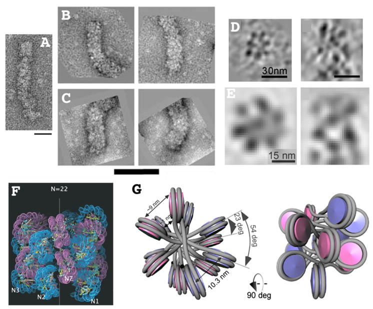
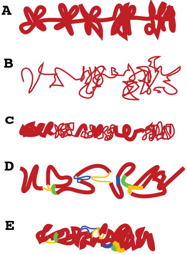
The past several years has seen increasing appreciation for plasticity of higher-level chromatin folding. Four distinct '30nm' chromatin fiber structures have been identified, while new in situ imaging approaches have questioned the universality of 30nm chromatin fibers as building blocks for chromosome folding in vivo. 3C-based approaches have provided a non-microscopic, genomic approach to investigating chromosome folding while uncovering a plethora of long-distance cis interactions difficult to accommodate in traditional hierarchical chromatin folding models. Recent microscopy based studies have suggested complex topologies co-existing within linear interphase chromosome structures. These results call for a reappraisal of traditional models of higher-level chromatin folding.


Figures

Figure 1
Figure 1. Four different higher-order chromatin structures and counting
(A) 21 nm diameter zig-zag structure formed after reconstitution with both core and linker histones using a 167 bp nucleosome repeat length. Reprinted from Fig. 4, reference [3], Copyright 2008 National Academy of Sciences, USA. (B-C) 33 nm diameter (B) versus 44 nm diameter (C) higher-order fibers formed after reconstitution with both core and linker histones but with nucleosome repeat lengths of 197 bp (B) or 207 (C). Reprinted from Fig. 1, reference [6], Copyright 2006 National Academy of Sciences, USA. (D-E) Transverse (left) or longitudinal (right) sections through individual (D) or averaged (E) tomograms of 32 nm diameter higher-order fibers visualized within cryo-sections of avian erythrocyte nuclei [9]. These fibers contain ~6.5 nucleosomes per 11 nm length of fiber. Reprinted from Figs. 1&2, reference [9]. (F) Molecular model [6] of one start helical structure proposed for 33 nm diameter reconstituted fiber (B). Inter-digitation of neighboring gyres of helix results in high nucleosome fiber density of ~ 11 nucleosomes per 11 nm fiber length. Reprinted from Fig. 3, reference [6], Copyright 2006 National Academy of Sciences, USA. (G) Molecular model for two-start, cross-linker type structure proposed for 32 nm fiber visualized by cryo-EM (D-E). The approximately 6.5 nucleosomes per 11 nm of fiber length corresponds more closely to previous estimates of fiber compaction. Reprinted from Fig. 3, reference [9], Copyright 2011 National Academy of Sciences, USA. Scale bars = 50 nm (A), 100 nm (B-C), 30 nm (D), or 15 nm (E).
Figure 2
Figure 2. Models for interphase chromosome large-scale chromatin folding
(A) Conventional model showing 30 nm higher order fibers forming clusters of loops. In older models these loop interactions are driven by poorly defined nuclear matrix interactions. This model is derived to a significant extent from extrapolation of radial loop models for mitotic chromosomes rather than direct visualization of interphase chromosomes. (B) Based on cryo-EM and ESI contrast for conventional EM, new models have suggested in vivo interphase chromosome structure consists nearly entirely of 10 nm fibers, locally dispersed or concentrated in compact local domains. (C) Chromonema fiber model, derived from a combination of light and electron microscopy, proposes existence of large-scale chromatin fibers on the order of 100 nm diameter. Irregular folding of 10 and/or 30 nm chromatin fibers underlies these large-scale fibers. Tight packing within these large-scale chromatin fibers has made it difficult to determine the substructure of these fibers and the relative ratio of 10 or 30 nm chromatin fibers. Discrete large-scale fiber segments are connected by less tightly coiled 10 and 30 nm fibers. (D) Chromatin hub model suggested by 3C experiments, in which looping interactions are formed through close interactions between various regulatory DNA elements. These loops are usually illustrated as composed of 30 nm fibers but could involve any combination of 10 and 30 nm fibers. (E) Hybrid chromonema / chromatin hub model in which complex topological looping interactions exist within large-scale chromatin fibers.

References

    1. Emanuel M, Radja NH, Henriksson A, Schiessel H. The physics behind the larger scale organization of DNA in eukaryotes. Physical biology. 2009;6:025008. - PubMed
    1. Grigoryev SA, Arya G, Correll S, Woodcock CL, Schlick T. Evidence for heteromorphic chromatin fibers from analysis of nucleosome interactions. Proceedings of the National Academy of Sciences of the United States of America. 2009;106:13317–13322. Combined experimental and theoretical approaches support the idea of features corresponding to both solenoid and zig-zag models for 30 nm higher order chromatin structure co-existing within single fibers as studied with reconstituted oligonucleosome arrays. A novel formaldehyde, electron microscopy based assay is used to examine preferential internucleosome interactions within these fibers which differ depending on the model. - PMC - PubMed
    1. Routh A, Sandin S, Rhodes D. Nucleosome repeat length and linker histone stoichiometry determine chromatin fiber structure. Proceedings of the National Academy of Sciences of the United States of America. 2008;105:8872–8877. - PMC - PubMed
    1. Schalch T, Duda S, Sargent DF, Richmond TJ. X-ray structure of a tetranucleosome and its implications for the chromatin fibre. Nature. 2005;436:138–141. - PubMed
    1. Dorigo B, Schalch T, Kulangara A, Duda S, Schroeder RR, Richmond TJ. Nucleosome arrays reveal the two-start organization of the chromatin fiber. Science. 2004;306:1571–1573. - PubMed

Publication types