Severe osteoporosis: diagnosis of femoral fractures
- PMID: 22460012
- PMCID: PMC3004454
Severe osteoporosis: diagnosis of femoral fractures
Abstract
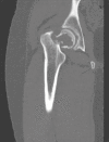
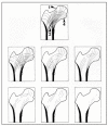
In the diagnosis of femoral fractures, Radiodiagnostic has a role in the different phases of the natural history of these lesions:- in diagnosis and characterization of fractures,- in follow up of the efficacy of therapy, evolution of fractures and any complications,- in studies of risk factors of fractures.Diagnostic imaging employs method of investigation as Conventional Radiology, still crucial in detection, characterization and control of fracture, Computed Tomography (CT) and Magnetic Resonance (MR), essential in doubt of occult fracture and in differential diagnosis between the possible causes of pathologic fracture. Finally, Dual X-ray Absorptiometry (DXA is still the fundamental methodic in diagnosis and assessment of osteoporosis, while QCT, pQCT and HR-CT are experimental techniques used to study in vivo bone microarchitecture and its metabolic and pathological changes.
Figures


Similar articles
-
Radiology of Osteoporosis.Can Assoc Radiol J. 2016 Feb;67(1):28-40. doi: 10.1016/j.carj.2015.02.002. Epub 2015 Jun 21. Can Assoc Radiol J. 2016. PMID: 26105503 Review.
-
Women with previous fragility fractures can be classified based on bone microarchitecture and finite element analysis measured with HR-pQCT.Osteoporos Int. 2013 May;24(5):1733-40. doi: 10.1007/s00198-012-2160-1. Epub 2012 Nov 20. Osteoporos Int. 2013. PMID: 23179565
-
Utilization of DXA Bone Mineral Densitometry in Ontario: An Evidence-Based Analysis.Ont Health Technol Assess Ser. 2006;6(20):1-180. Epub 2006 Nov 1. Ont Health Technol Assess Ser. 2006. PMID: 23074491 Free PMC article.
-
[Imaging of diabetic osteopathy].Radiologe. 2015 Apr;55(4):329-36. doi: 10.1007/s00117-014-2723-6. Radiologe. 2015. PMID: 25895468 German.
-
Use of dual energy X-ray absorptiometry, the trabecular bone score and quantitative computed tomography in the evaluation of chronic kidney disease-mineral and bone disorders.Nephrology (Carlton). 2017 Mar;22 Suppl 2:19-21. doi: 10.1111/nep.13016. Nephrology (Carlton). 2017. PMID: 28429557 Review.
Cited by
-
Osteoprotective Activity and Metabolite Fingerprint via UPLC/MS and GC/MS of Lepidium sativum in Ovariectomized Rats.Nutrients. 2020 Jul 13;12(7):2075. doi: 10.3390/nu12072075. Nutrients. 2020. PMID: 32668691 Free PMC article.
-
Age as a Risk Factor for Intraoperative Periprosthetic Femoral Fractures in Cementless Hip Hemiarthroplasty for Femoral Neck Fractures: A Retrospective Analysis.Clin Orthop Surg. 2024 Feb;16(1):41-48. doi: 10.4055/cios23157. Epub 2023 Dec 20. Clin Orthop Surg. 2024. PMID: 38304210 Free PMC article.
-
The Impact of Visual Input and Support Area Manipulation on Postural Control in Subjects after Osteoporotic Vertebral Fracture.Entropy (Basel). 2021 Mar 20;23(3):375. doi: 10.3390/e23030375. Entropy (Basel). 2021. PMID: 33804770 Free PMC article.
References
-
- Rossigni M, Piscitelli P, Fitto F, et al. Incidenza e costi delle fratture di femore in Italia. Incidence and socioeconomic burden of hip fractures in Italy. Reumatismo. 2005;57(2):97–102. - PubMed
-
- Hossain M, Barwick C, Sinha AK, et al. Is magnetic resonance imaging (MRI) necessary to exclude occult hip fracture? Injury. 2007 Oct;38:1204–8. - PubMed
-
- Sankey RA, Turner J, Lee J, et al. The use of MRI to detect occult fractures of the proximal femur: a study of 10 consecutive cases over a ten-year period. J Bone Joint Surg Br. 2009 Aug;91(8):1064–8. - PubMed
-
- Dupuis MG, Moussaoui A, Douzal V, et al. Imaging of traumatic injuries of the hip. J Radiol. 2007 May;88(5 Pt 2):760–74. - PubMed
-
- Hong RJ, Hughes TH, Gentili A, et al. Magnetic resonance imaging of the hip. J Magn Reson Imaging. 2008 Mar;27(3):435–45. - PubMed
LinkOut - more resources
Full Text Sources
