

Definition of bone necrosis by the pathologist
- PMID: 22460748
- PMCID: PMC2781178
Definition of bone necrosis by the pathologist
Abstract
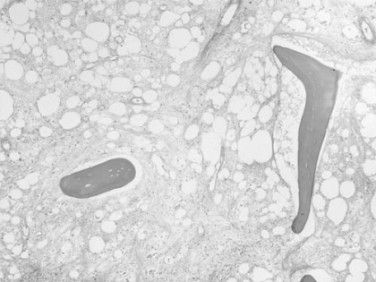
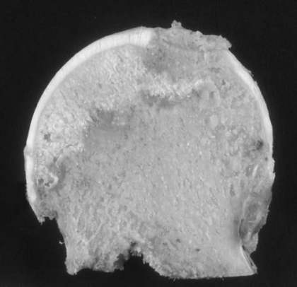
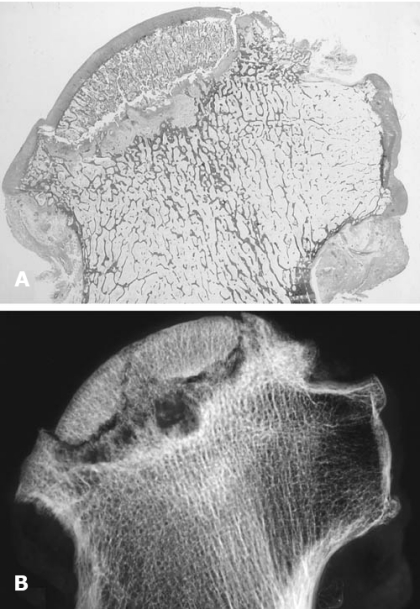
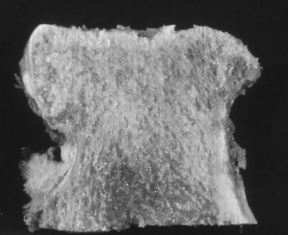
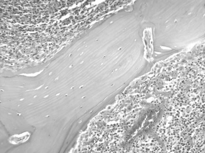
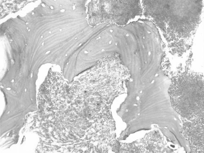
Osteonecrosis is a common disorder that may go clinically unrecognized or may result in the collapse of the architecture of bone, determining severe anatomic alterations of the involved site. Osteonecrosis is not a specific disease entity, but rather the result of a number of conditions ultimately leading to an impairment of blood supply to the bone tissue, although there is evidence that modifications of bone remodelling activity and weakening of bone structure with formation of microfractures are implicated as well. According to the site involved and to the factors promoting its development, the morbid anatomy and histopathology of osteonecrosis show a different appearance. This review discusses the main skeletal manifestations of osteonecrosis, including subarticular avascular necrosis of the femoral head and of the knee, as well as osteonecrosis of the jaw.
Figures
References
-
- McCarthy EF. Aseptic necrosis of bone. An historic perspective. Clin Orthop Relat Res. 1982;168:216–221. - PubMed
-
- Bonfiglio M. Aseptic necrosis of the femoral head in dogs. Effects of drilling and bone grafting. Surg Gynecol Obstet. 1954;98:591–599. - PubMed
-
- Catto M. A histological study of avascular necrosis of the femoral head after transcervical fracture. J Bone Joint Surg. 1965;47B:749–776. - PubMed
-
- Young MH. Epiphyseal infarction in a growing long bone. An experimental study in the rabbit. J Bone Joint Surg. 1966;48B:826–840. - PubMed
-
- Frost HM. In vivo osteocyte death. J Bone Joint Surg. 1960;42A:138–143. - PubMed
LinkOut - more resources
Full Text Sources
Other Literature Sources